|
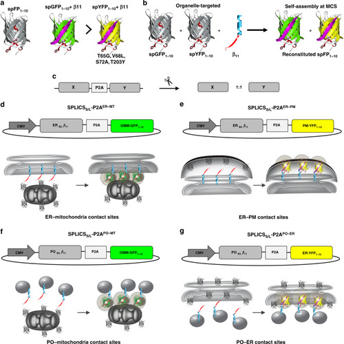
Design of the SPLICS<sub>S/L</sub>-P2A probes.a Cartoon representation of GFP/YFP SPLICSS/L-P2A probes. The cartoon also indicates the occurrence of reconstitution of the two spectral variants through association with the invariant strand β11. b Schematic representation of the targeted GFP/YFP SPLICSS/L-P2A chimeras. GFP/YFP protein was split in two non-fluorescent portions, G(Y)FP1–10 and β11 fragment that are targeted to outer face of organelles of interest. The complementation of targeted split-GFP at membrane contact sites was also shown in the cartoon. c Creation of a single vector for the equimolar expression of the targeted fragments through the insertion of the P2A peptide. d–g Schematic representations of the SPLICSS/L-P2A vectors. The β11S/L coding sequence is cloned upstream the viral P2A peptide sequence, while the second cassette is occupied by the G(Y)FP1–10. d SPLICSS/L-P2AER–MT expression plasmids (ERS/L-β11 and OMM-GFP1–10). e SPLICSS/L-P2AER–PM expression plasmids (ERS/L-β11 and PM-YFP1–10). f SPLICSS/L-P2APO–MT expression plasmids (POS/L-β11 and OMM-GFP1–10). g SPLICSS/L-P2APO–ER expression plasmids (POS/L-β11 and ER-YFP1–10).
|