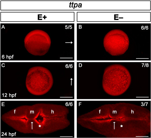
Ttpa signal localized throughout early embryo and brain ventricle borders regardless of VitE status. Ttpa expression in E+ and E− embryos is indicated with red fluorescence; dorsal direction is indicated by arrow (A–D). At 6 hpf (dorsal shield stage, A, B), Ttpa expression was present throughout the animal poles [E+ embryos, n = 5/5 (n = number of animals with the observed defect/total number of animals observed)], E− embryos, n = 6/6). At 12 hpf (90% epiboly, C, D), ttpa expression was present both in the embryo and in the yolk syncytial layer (E+ embryos, n = 6/6; E− embryos, n = 7/8 ), arrow indicates anterior region of the embryo. At 24 hpf (E, F), Ttpa expression was localized in the brain ventricle borders and within cells of the fore (f), mid- (m), and hindbrain (h). Arrows indicate the midbrain-hindbrain boundary where diencephalic ventricle expansion was altered; *Represent inflation in E+ embryos (E, n = 6/6) or lack thereof in E− embryos (F, n = 3/7). Scale bar represents 500 μm (A–D) and 50 μm (E, F); representative embryos are shown. Figure panels were generated with the BZ- × 700 microscope, processed with BZ-X Analyzer Software with image adjustments equally applied across time points in Adobe Photoshop v21.2.1.
|
