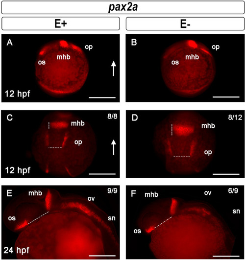
Midbrain-hindbrain boundary formation shown by pax2a expression is dysregulated by VitE deficiency. Pax2a expression in early optic stalk (os), midbrain-hindbrain boundary (mhb) and otic placode (op) in 12 hpf embryos, lateral views (A, B); arrow indicates dorsal region. At 12 hpf, E+ embryos (C) had defined mhb and op borders (n = 8/8), while E− embryos (D) had diffuse mhb and op borders (n = 8/12 assessed). Shown are representative E+ embryo with mhb 25 μm wide and op 49 μm apart, while representative E− embryo measurements were mhb 42 μm wide and op 63 μm apart. At 24 hpf, in E+ (E) and E− embryos (F) pax2a was expressed in the os, mhb, otic vesicles (ov) and spinal cord neurons (sn). Distance between os and mhb, a measure of first brain ventricle inflation, were greater in a representative E+ (91 μm) relative to an E− (80 μm) embryo. E+ embryo (E) spinal cord neurons at the same fluorescence exposure had significantly increased pax2a signal (n = 9/9), as compared with E− embryos (n = 6/9). Scale bar represents 100 μm; representative embryos are shown. Figure panels were generated with the BZ- × 700 microscope, processed with BZ-X Analyzer Software with image adjustments equally applied across time points in Adobe Photoshop v21.2.1.
|
