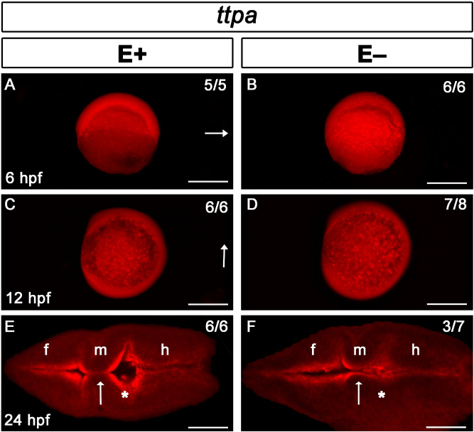
Ttpa signal localized throughout early embryo and brain ventricle borders regardless of VitE status. Ttpa expression in E+ and E− embryos is indicated with red fluorescence; dorsal direction is indicated by arrow (A–D). At 6 hpf (dorsal shield stage, A, B), Ttpa expression was present throughout the animal poles [E+ embryos, n = 5/5 (n = number of animals with the observed defect/total number of animals observed)], E− embryos, n = 6/6). At 12 hpf (90% epiboly, C, D), ttpa expression was present both in the embryo and in the yolk syncytial layer (E+ embryos, n = 6/6; E− embryos, n = 7/8 ), arrow indicates anterior region of the embryo. At 24 hpf (E, F), Ttpa expression was localized in the brain ventricle borders and within cells of the fore (f), mid- (m), and hindbrain (h). Arrows indicate the midbrain-hindbrain boundary where diencephalic ventricle expansion was altered; *Represent inflation in E+ embryos (E, n = 6/6) or lack thereof in E− embryos (F, n = 3/7). Scale bar represents 500 μm (A–D) and 50 μm (E, F); representative embryos are shown. Figure panels were generated with the BZ- × 700 microscope, processed with BZ-X Analyzer Software with image adjustments equally applied across time points in Adobe Photoshop v21.2.1.
This image is the copyrighted work of the attributed author or publisher, and
ZFIN has permission only to display this image to its users.
Additional permissions should be obtained from the applicable author or publisher of the image.
Full text @ Sci. Rep.
Your Input Welcome
Thank you for submitting comments. Your input has been emailed to ZFIN curators who may contact you if
additional information is required.
Oops. Something went wrong. Please try again later.