
Fig. 6-S4
- ID
- ZDB-FIG-190201-18
- Publication
- Naylor et al., 2018 - A novel mechanism of gland formation in zebrafish involving transdifferentiation of renal epithelial cells and live cell extrusion
- Other Figures
- All Figure Page
- Back to All Figure Page
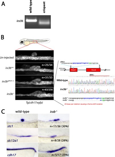
irx3b crispants and stable mutants recapitulate the renal phenotypes associated with irx3b morpholino knock down. (A) Panel shows the gel image of the irx3b PCR product amplified from genomic DNA and processed through the T7 endonuclease one protocol (see materials and methods section). (B) Panels show lateral views of Tg(cdh17:egfp) embryos in wild-type, irx3b morphant, irx3b crispant and germline stable irx3b mutant embryos. Sanger sequencing analysis (right) shows a 29 bp deletion that causes a frame-shift mutation in exon 1 of the irx3b gene. (C) Panels show lateral views of the posterior trunk of stable irx3b mutants stained for stc1, slc12a1 and cdh17 transcripts. |
| Genes: | |
|---|---|
| Fish: | |
| Anatomical Terms: | |
| Stage: | Long-pec |
| Fish: | |
|---|---|
| Observed In: | |
| Stage: | Long-pec |