
FIGURE
Fig. 2-S2
- ID
- ZDB-FIG-190201-8
- Publication
- Naylor et al., 2018 - A novel mechanism of gland formation in zebrafish involving transdifferentiation of renal epithelial cells and live cell extrusion
- Other Figures
- All Figure Page
- Back to All Figure Page
Fig. 2-S2
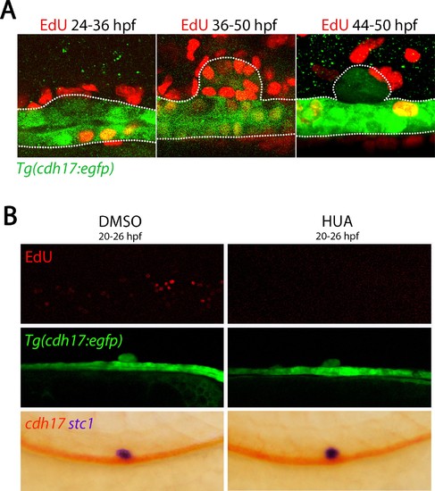
Cell division is not required for CS formation. (A) Panels show lateral views of Tg(cdh17:egfp) embryos stained for EdU labelling at 36 hpf (left panel) and 50 hpf (two right panels) after treatment with EdU between the stages shown. (B) Lateral views of DMSO vehicle control (left panels) and HUA treated animals treated from 20 to 26 hpf and then fixed at 50 hpf. Top panels are EdU labelling, middle panels are of Tg(cdh17:egfp) embryos and bottom panels are of embryos stained for cdh17 (red) and stc1 (purple) expression. |
Expression Data
Expression Detail
Antibody Labeling
Phenotype Data
Phenotype Detail
Acknowledgments
This image is the copyrighted work of the attributed author or publisher, and
ZFIN has permission only to display this image to its users.
Additional permissions should be obtained from the applicable author or publisher of the image.
Full text @ Elife