
FIGURE
Fig. 6-S2
- ID
- ZDB-FIG-190201-16
- Publication
- Naylor et al., 2018 - A novel mechanism of gland formation in zebrafish involving transdifferentiation of renal epithelial cells and live cell extrusion
- Other Figures
- All Figure Page
- Back to All Figure Page
Fig. 6-S2
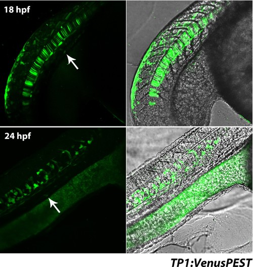
Notch activity in the pronephros is not observed early in development in the TP1:VenusPEST reporter line. Top panels show lateral views of the trunk of an 18 hpf embryo, bottom panels show lateral views of the trunk region at 24 hpf. Venus+ cells in the region of the pronephros (arrows) were in the epidermis when analysed along the Z-axis. |
Expression Data
Expression Detail
Antibody Labeling
Phenotype Data
Phenotype Detail
Acknowledgments
This image is the copyrighted work of the attributed author or publisher, and
ZFIN has permission only to display this image to its users.
Additional permissions should be obtained from the applicable author or publisher of the image.
Full text @ Elife