
Fig. 3
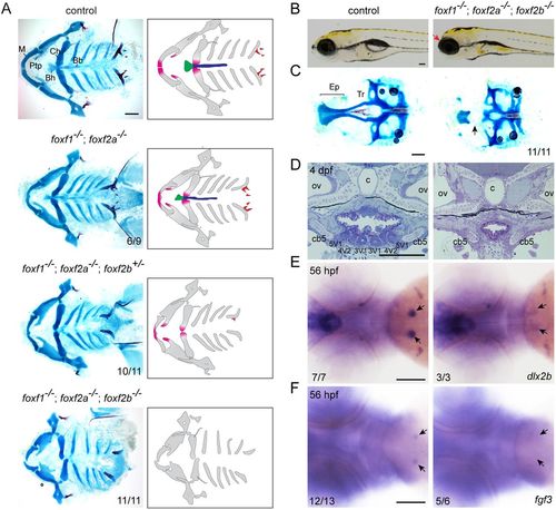
Loss of midline cartilage and pharyngeal teeth in Fox-F mutants. (A) Ventral views of dissected facial skeletons, with cartilage in blue and bones and teeth in red. Schematics show affected elements in color. Bb, basibranchials; Bh, basihyal; Ch, ceratohyal; M, Meckel's; Ptp, pterygoid process. (B) Lateral views of wild-type sibling control and foxf1; foxf2a; foxf2b triple mutant at 5 dpf. Mutants show anterior truncation of the head (arrow). (C) Dissections of neurocranial cartilages show loss of the trabecular cartilages (Tr, arrow) in foxf1; foxf2a; foxf2b triple mutants. Ep, ethmoid plate. (D) Sagittal sections through the tooth-forming seventh arch of a wild-type sibling control and foxf1; foxf2a; foxf2b triple mutant at 4 dpf. All teeth (3V1, 4V2 and 5V1) are missing in mutants. c, chorda; cb5, ceratobranchial 5; ov, otic vesicle. (E,F) Whole-mount in situ hybridizations show loss of dlx2b (E) and fgf3 (F) in the developing tooth regions (arrows) of foxf1; foxf2a; foxf2b triple mutants. Numbers denote proportions of embryos with displayed phenotypes or expression patterns. Scale bars: 25 μm. |
| Genes: | |
|---|---|
| Fish: | |
| Anatomical Term: | |
| Stage: | Long-pec |