
Fig. 2
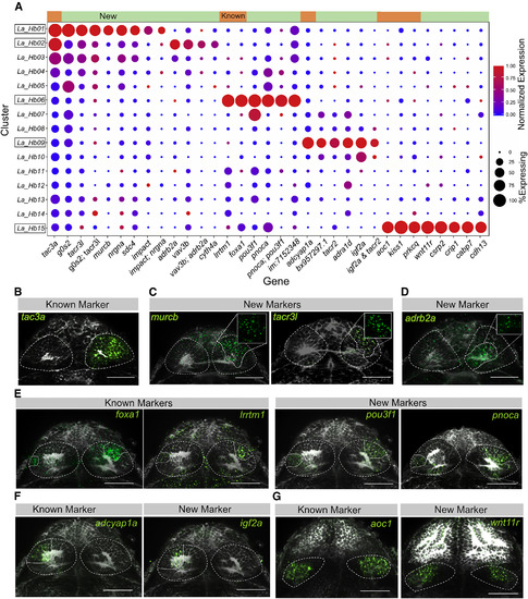
Validation and Spatial Distribution of Previously Described Neuronal Types along with Identified Novel Markers (A) Expression profiles of known and novel habenular marker genes that are specific or enriched in the five clusters displaying previously described gene expression signatures. Green bar on top represents new markers, and orange bar represents known markers. (B–G) In vivo expression patterns of known and novel marker genes that are enriched in clusters harboring previously characterized habenular genes (Hb01, Hb02, Hb06, Hb09, and Hb15). Each type was characterized by both previously described markers, and new markers found from single-cell analysis. RNA-FISH (green) was performed with a total-Erk (pale gray) co-stain for registration (see Figure 3). In some cases, a non-linear filter (gamma = 0.3) was applied to the total-Erk (gray) channel to aid visualization of the in situ signal (green). (B–D) FISH labeling of (B) previously known marker (tac3a) and new markers for (C) Hb01 (murcb, tacr3l) and (D) Hb02 (adrb2a) found by single-cell analysis. Insets show regionalized expression of the gene without total-Erk. (E) FISH labeling of new markers pou3f1 and pnoca enriched in the lrrtm1+ and foxa1+ cluster Hb06. (F) FISH labeling of new marker igf2a enriched in adcyap1a+ left-only cluster Hb09. (G) FISH labeling of new marker wnt11r specific to the aoc1+ ventral habenular cluster Hb15. Scale bars indicate 50 μm. See also Figures S2 and S3, Table S1, and Movie S1. |
| Genes: | |
|---|---|
| Fish: | |
| Anatomical Terms: | |
| Stage: | Days 7-13 |