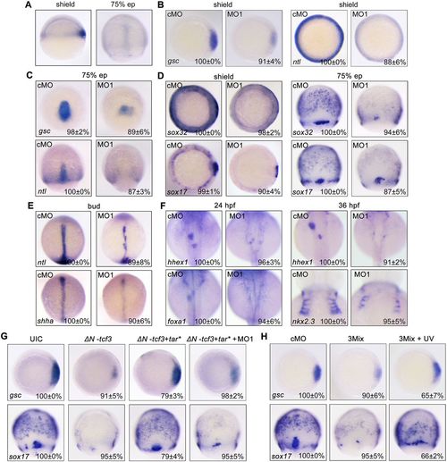
net1 is crucial for mesendoderm formation in zebrafish embryos. (A) Visualization of net1 expression in zebrafish embryos at shield and 75% epiboly (ep) stages using whole-mount in situ hybridization. Shield stage, lateral views with dorsal to the right; 75% epiboly stage, dorsal views with animal pole to the top. (B–F) The expression of mesendoderm marker genes and mesendoderm derivative marker genes in cMO- and net1 MO1-injected embryos at the indicated stages. (B–D) Shield stage, animal views with dorsal to the right; 75% epiboly stage, dorsal views with animal pole to the top. (E,F) Bud stage, and 24 and 36 hpf. All images are shown in dorsal views with anterior to the top. (G) Wild-type embryos were injected with 50 pg ΔN-tcf3 mRNA alone or co-injected with 3 pg tar* mRNA and 4 ng net1 MO1, and then collected at the shield and 75% epiboly stages for whole-mount in situ hybridization with a gsc or sox17 probe. (H) Overexpression of net1 rescues mesendoderm induction defects in net1 morphants. Wild-type embryos were injected with cMO or net1 3Mix (4 ng net1 MO1, 200 pg Flag-net1 mRNA and 1 ng AS-Flag-photo-MO), and then a subset of net1 3Mix-injected embryos were exposed to UV light at the sphere stage. The expression of gsc and sox17 was examined by in situ hybridization at shield and 75% epiboly stages, respectively. In B–H, the percentages (mean±s.d.) of the affected embryos are shown as calculated from three independent biological repeats with ∼15–30 embryos in each group. UIC, uninjected control.
|
