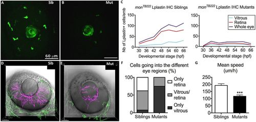
Moonshine macrophages display a defective recruitment towards the eye and into the retina. (A,B) In vivo observation of monNQ039Tg(Pu1:gfp) siblings (Sib) (A) and mutants (Mut) (B) at 30 hpf reveals that several GFP-positive myeloid cells have already arrived at the eye in the siblings, whereas there are few in the mutants; the lens shows apparent green fluorescence only due to its optical properties. (C) Counts (Nb, number) of L-plastin-positive leukocytes in the retina (red curve) and in the vitreous space (from where wt macrophages enter the retina; light blue curve) of monTB222/Tü siblings (left graph, 10–18 embryos/timepoint) and mutants (right graph, 10–16 embryos/timepoint) show an increasing number of macrophages in the eye (blue curve) and resulting colonization of the retina (red curve) in the sibling, whereas very few macrophages arrive at the eye in the mutants and their number does not increase with time. IHC, immunohistochemistry. (D–G) 4D tracking for 12 h of GFP-positive myeloid cells inside (pink tracks) and outside (green tracks) the eye in monNQ039Tg(Pu1:gfp) siblings (D) and mutants (E) from 48 hpf showing that the few mutant cells inside the eye mostly do not invade the retina (n=20 cells from two embryos), whereas it is the opposite in the siblings (n=66 cells from two embryos), as quantified in F. (G) Myeloid cells in the mesenchyme peripheral to the eye (green tracks) migrate more slowly in the mutants (n=42 cells from two embryos, mean speed 116.7 µm/h) than in the siblings (n=109 cells from two embryos, mean speed 192.9 µm/h). Error bars show the s.e.m. ***P<0.001.
|
