
Fig. S2
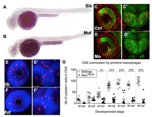
Macrophages do not colonize the CNS in monTB222 mutants and Trim33 morphants. (A-B) Csfr1a ISH at 36 hpf shows that the initial primitive macrophage production and spreading in the monTB22 mutant (B) are similar to the sibling (A). (C-D') In vivo images of retinas (C,D) or midbrain (C'-D') of 3 days old Tg(HuC:GFP; mpeg1:Gal4; UAS:NfsB-mCherry) embryos injected (D-D') or not (C,C') with the Trim33 morpholino. Trim33 KO (D,D') causes macrophage depletion in both CNS compartments. (E-G) Lplastin whole-mount immunodetection (combined with DAPI detection of cell nuclei) in monTB222/Tü siblings (E,E') and mutants (F,F') reveals numerous leukocytes (macrophages) in the retinas (E) and midbrain (E') of 3 days old siblings, whereas there are only a few in the mutants. Counting of these cells every 6 hrs from 30 to 66 hpf reveals that monTB222 mutants have less leukocytes in the CNS at all stages. Ctrl, control; Mo, morphant; Mut, mutant; nb, number; Sib, sibling. **, P value <0.01; ***, P value < 0.001. |
| Gene: | |
|---|---|
| Fish: | |
| Anatomical Term: | |
| Stage: | Prim-25 |