
Fig. S2
- ID
- ZDB-FIG-161221-41
- Publication
- Chen et al., 2016 - MicroRNA-126a Directs Lymphangiogenesis Through Interacting With Chemokine and Flt4 Signaling in Zebrafish
- Other Figures
- All Figure Page
- Back to All Figure Page
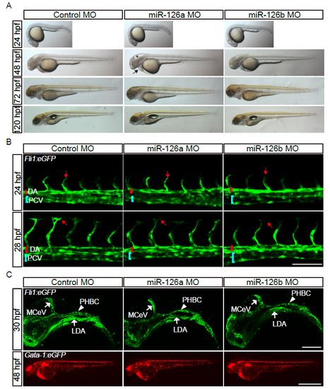
The gross morphology and the anatomic structure of early blood vessels are normal in miR-126a morphants. (A) Bright-field images of embryos injected with control MO (2 pmol), miR-126a MO (2 pmol) and miR-126b MO (2 pmol) showing normal gross morphology. Asterisk indicates brain development defects and arrow denotes pericardial edema. (B) Confocal images of trunk blood vascular in 24 and 28 hpf embryos illustrating normal angiogenesis. DA indicates dorsal aorta and PCV denotes posterior cardinal vein. The red arrow indicates migration of the tip cell. Scale bar, 100 μm. (C) Confocal images of head blood vascular in 30 hpf embryos, scale bar is 50 μm (Up panels). Stereomicroscope images of blood circulation in 48 hpf embryos, scale bar is 100 μm (Bottom panels). |