
Fig. S1
- ID
- ZDB-FIG-161221-40
- Publication
- Chen et al., 2016 - MicroRNA-126a Directs Lymphangiogenesis Through Interacting With Chemokine and Flt4 Signaling in Zebrafish
- Other Figures
- All Figure Page
- Back to All Figure Page
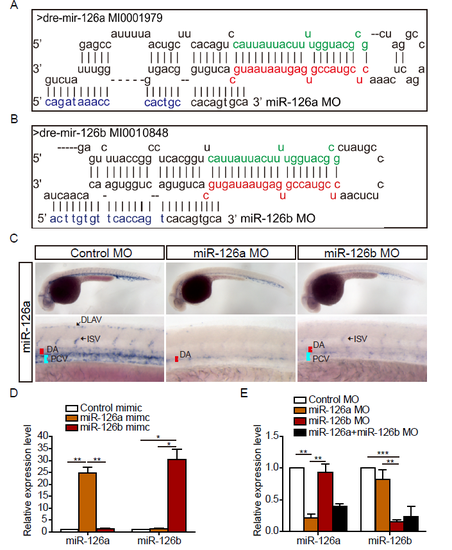
miR-126a and miR-126b could be specifically knocked down by morpholino. (A and B) Schematic alignment of miR-126a MO and miR-126b MO with pri-miR-126a and pri-miR-126b. The letters in green denote miR-126a* (miR-126a-5p) and miR-126b* (miR-126b-5p), respectively. The red letters indicate miR-126a (miR-126a-3p) and miR-126b (miR-126b-3p), respectively. The blue letters indicate the different nucleotides between miR-126a MO and miR-126b MO. (C) Whole-mount in situ hybridization (WISH) for miR-126a at 36 hpf embryos. DA: dorsal aorta; PCV: posterior cardinal vein; DLAV: dorsal longitudinal anatomic vessel; ISV: intersegmental vessel. (D) Quantification of mature miR-126a and miR-126b in 72 hpf embryos injected with mimics (0.4 pmol) indicating miR-126a and miR-126b could be specifically detected by real-time PCR. (E) Quantification of mature miR-126a and miR-126b in 72 hpf embryos injected with MOs (2 pmol of single MO, 1 pmol of each MO when co-injected). ***p<0.001, **p<0.001, *p<0.05. |