
Fig. 3
- ID
- ZDB-FIG-161221-36
- Publication
- Chen et al., 2016 - MicroRNA-126a Directs Lymphangiogenesis Through Interacting With Chemokine and Flt4 Signaling in Zebrafish
- Other Figures
- All Figure Page
- Back to All Figure Page
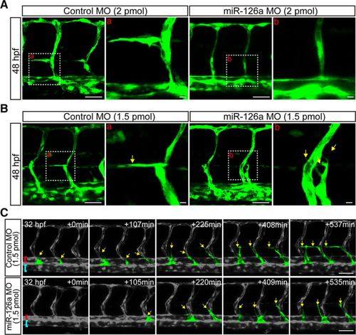
Deficiency of miR-126a blocks parachordal lymphangiblast (PL) extension along horizontal myoseptum. A, Confocal images of trunk vessels at 48 hours post fertilization (hpf) embryos showing no lymphangiogenic sprouts in miR-126a morphants (2 pmol). Scale bars, 40 μm. B, Confocal images of trunk vessels in 48 hpf embryos injected with control morpholino (MO; 1.5 pmol) or miR-126a MO (1.5 pmol) revealing the extension of PL was blocked by miR-126a knockdown. Scale bars, 40 μm. C, Still images from time-lapse analysis of control and miR-126aKD embryos. Time (min, begin at 32 hpf) is noted in the top right corner. Lateral views, dorsal is up, anterior to the left. Yellow arrows indicate the lymphatic endothelial cells (LECs) emerging from posterior cardinal vein (PCV). The extension along horizontal myoseptum of LECs was normal in control embryos, but this process was blocked in miR-126aKD embryos. Red brackets, dorsal aorta (DA); blue rackets, PCV. Scale bars, 50 μm. |
| Fish: | |
|---|---|
| Knockdown Reagent: | |
| Observed In: | |
| Stage: | Long-pec |