|
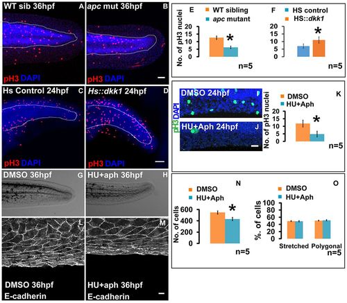
Patterning in the median fin epithelium is not influenced by proliferation rates and total cell numbers. Phospho-histone 3 and DAPI staining under given genetic conditions or treatments at 24 hpf (A-D,I,J). (E,F,K) Quantification reveals decrease in proliferation in apc mutant (E) and drug (HU+aph) treated (K) embryos, and an increase in HS:dkk1-GFP embryos (F) when compared with their respective controls. (G,H) Median fin morphology at 36 hpf in DMSO- (G) and HU+aph-treated embryos (H). (L,M) Cellular morphology in 36 hpf control (L) and HU+aph-treated (M) embryos. (N) Comparison of total number of cells in the fin fold of DMSO control and HU+aph-treated embryos. Proportions of stretched and polygonal cells in control and HU+aph-treated embryos at 36 hpf (O). Scale bars: 50 μm in A-D,I,J; 10 μm in L,M. *P<0.01 (Student′s t-test).
|