|
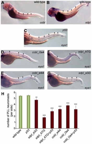
Knock-down of either cobl or syndapin I diminishes neuromasts of the posterior lateral line organ. (A,B) Wild-type zebrafish (72hpf) analyzed for cobl and syndapin I expression patterns by in situ hybridization. Arrowheads mark regular arrays of cobl- and syndapin I-positive spots in the tails. (C–G) In situ hybridization of eya1 (neuromast marker) at 48hpf. Neuromasts at the facing tail side are marked by arrowheads. (D–G) Knock-down of cobl (D,E) and syndapin I (F,G) leads to reduced neuromast numbers. (H) Quantitative analysis. Wild-type, n = 117; MO p53, n = 55; MO sdpI_e3i3, n = 27; MO sdpI_ATG, n = 58; MOs sdpI_ATG+p53, n = 17; MO cobl_e4i4, n = 16; MO cobl_i3e4, n = 48; MOs cobl_i3e4+p53, n = 26. ANOVA with Tukey’s test versus MO p53; **P<0.01 for MO sdpI_e3i3 and ***P<0.001 for all other data sets. PLL, posterior lateral line.
|