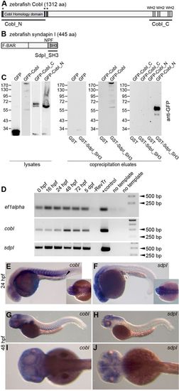
Biochemical interaction and expression of zebrafish Cobl and syndapin I. (A) Domain structure of zebrafish Cobl (JQ776648) with Cobl homology domain, three KrRAPpPP motifs (asterisks) and three WH2 domains. Black lines represent deletion mutants used in the present study. (B) Domain structure of zebrafish syndapin I and a deletion mutant used. NPF, Asn-Pro-Phe motif. (C) Co-precipitation experiments with zebrafish Cobl and N- and C-terminal fragments thereof (Cobl_N, Cobl_C) expressed as GFP-fusion proteins and immobilized, recombinant GST-syndapin I SH3 domain (GST-SdpI_SH3) and GST, respectively, analyzed by anti-GFP immunoblotting of cell lysates (input) and eluates. (D) Expression levels of cobl and syndapin I (sdpI) at 0hpf to 5dpf analyzed by RT-PCR. –RevTr, without reverse transcriptase. (E–J) Tissue-specific expression patterns of cobl (E,G,I) and syndapin I (F,H,J) analyzed by in situ hybridization at 24hpf (E, F) and 48hpf (G–J). Insets in E and F show dorsal head views. Cobl and syndapin I are expressed in neuronal tissues. Cobl expression was additionally detected in somites (especially at 24hpf; E) and the pronephros (arrowhead in E).
|
