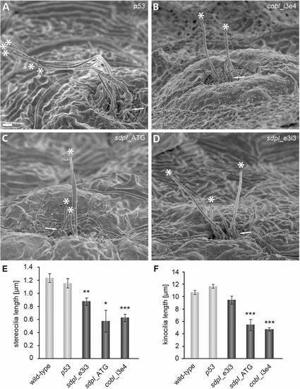
Both F-actin-rich stereocilia and microtubule-based kinocilia are defective in cobl and sdpI morphants. (A–D) SEM images of hair cells of neuromasts of zebrafish embryos injected as indicated at 72hpf. In both cobl- (B) and sdpI-morphant fish (C,D), kinocilia are reduced in number and significantly shortened. Tips of kinocilia are marked by asterisks. F-actin-rich stereocilia (arrows in A–D mark examples) also are severely affected. Scale bar: 1μm. (E,F) Quantitative analyses of observable stereocilia (E) and kinocilia (F). Wild-type, n = 99 kinocilia/82 stereocilia; control MO p53, n = 80 kinocilia/17 stereocilia; MO sdpI_e3i3, n = 37 kinocilia/38 stereocilia; MO sdpI_ATG, n = 10 kinocilia/5 stereocilia; MO cobl_i3e4, n = 58 kinocilia/19 stereocilia. Statistical analyses were performed versus wild-type. *P<0.05, **P<0.01, ***P<0.001. Kinocilia defects observed in cobl and syndapin I morphants (F) show even higher statistical significances versus MO p53-control (not depicted), MO sdpI_e3i3 (**), MO sdpI_ATG (***) and MO cobl_i3e4 (***).
|
