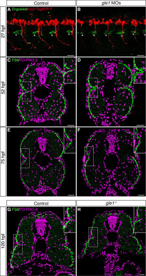
The integrity of the horizontal myoseptum in gle1 morphants and mutants appears intact. (A,B) Confocal images of control (A) and gle1 morphant (gle1 MOs) (B) zebrafish embryos with motoneurons labeled by mnx1:TagRFP-T transgene and muscle pioneers (MPs) labeled by anti-Engrailed/4D9 antibody (1:100 dilution) at 27 hpf. There were no discernible differences in the number and organization of the Engrailed+ MPs (asterisks) around the horizontal myoseptum between the control and gle1 morphant (<4-6 MPs per side in each somite segment). (C-H) Transverse cryosections through the trunk of controls (C,E,G), gle1 morphants (D,F) and gle1-/- mutant (H) were stained with anti-F59 antibody (1:100 dilution) to reveal the slow muscle fibers beneath the skin at 52 hpf (C,D), 75 hpf (E,F) and 120 hpf (G,H). At all three stages examined, the organization of the muscle fibers around the horizontal myoseptum (e.g. boxed areas, enlarged in insets) appeared indistinguishable between the controls and gle1 morphants/mutants. Nuclei were counterstained by TO-PRO-3. Scale bars: 25 μm.
|
