
Fig. S2
- ID
- ZDB-FIG-120405-23
- Publication
- Jao et al., 2012 - A zebrafish model of lethal congenital contracture syndrome 1 reveals Gle1 function in spinal neural precursor survival and motor axon arborization
- Other Figures
- All Figure Page
- Back to All Figure Page
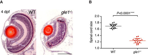
gle1-/- mutants have small eyes and spinal cord. (A) H&E-stained frontal transverse sections through the eyes of wild-type (WT) and gle1-/- mutant larvae at 4 dpf. All retinal cell layers are present in the gle1-/- eye, albeit in reduced numbers. Cells with condensed nuclei typical of apoptotic cells can be seen in the gle1-/- retina, especially in the ciliary marginal zone (red dashed outlines). cmz, ciliary marginal zone; prl, photoreceptor layer; opl, outer plexiform layer; onl, outer nuclear layer; inl, inner nuclear layer; ipl, inner plexiform layer; rgc, retinal ganglion cell layer; le, lens. Images were taken under the same magnification. Scale bar: 100 μm. (B) Quantification of spinal cord size (representative spinal cord sections shown in Fig. 2K,L) by measuring the area of 7-μm transverse plastic sections of the spinal cord at the region above the yolk extension using ImageJ. Data were collected from three randomly selected 5-dpf gle1-/- mutant larvae and three of their wild-type siblings; six sections per larva were measured. Statistical significance was determined using the unpaired t-test. The spinal cords of the gle1-/- mutant larvae were approximately 73% of wild-type size. |
| Fish: | |
|---|---|
| Observed In: | |
| Stage: | Day 4 |