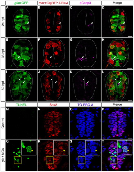
Neural precursors, rather than differentiated neurons, undergo apoptosis in gle1 morphants. (A-L) Transverse cryosections through the spinal cords of gle1 morphants harboring gfap:GFP and mnx1:TagRFP-T transgenes were stained with anti-Elavl and anti-cleaved caspase 3 (aCasp3) antibodies at 24 (A-D), 36 (E-H) and 52 (I-L) hpf. The gfap:GFP transgene reveals the radial glia (A,E,I); anti-Elavl staining reveals newly differentiated neurons with a cytoplasmic signal; the mnx1:TagRFP-T transgene labels motoneurons with both nuclear and cytoplasmic signals (B,F,J); aCasp3 labels apoptotic cells (C,G,K). aCasp3+ cells are often gfap:GFP+ (arrows), located close to the central canal (dashed lines), and/or in close proximity with Elavl+ mnx1:TagRFP-T+ neurons. Elavl+ mnx1:TagRFP-T+ neurons are always negative for aCasp3 (merged channels, D,H,L). (M-T) Transverse cryosections through the spinal cords of control (M-P) and gle1 morphant (Q-T) embryos at 31 hpf were stained for Sox2, followed by TUNEL reactions, to reveal Sox2+ neural precursors (N,R) and TUNEL+ apoptotic cells (M,Q). Nuclei were counterstained with TO-PRO-3 (O,S). Detection of Sox2+ TUNEL+ nuclei (arrowheads) indicates that Sox2+ neural precursors die upon Gle1 depletion. Each image is a single optical section (<1 μm) extracted from the confocal z-stack (dorsal to top). Dashed lines outline spinal cords and central canals. Scale bars: 10 μm in D,H,L; 7.5 μm in P,T.
|
