
Fig. 2
- ID
- ZDB-FIG-120209-18
- Publication
- Zhang et al., 2012 - Zebrafish Models for Dyskeratosis Congenita Reveal Critical Roles of p53 Activation Contributing to Hematopoietic Defects through RNA Processing
- Other Figures
- All Figure Page
- Back to All Figure Page
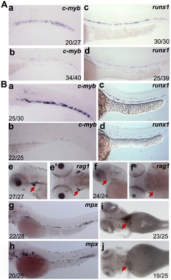
Analysis of hematopoietic defects in dkc1 and nola1 deficiency. (A) Expression of HSC marker genes was decreased in dkc1 morphants. C-myb expression at 3 dpf (a and b); runx1 expression at 30 hpf (c and d). (B) Number of HSC (a, b, c and d) and red blood cells (red arrow in i and j) at 4 dpf were significantly reduced in nola1 mutants. C-myb expression at 3 dpf; runx1 expression at 30 hpf. Granulocytes, marked by mpx, weren′t affected or slightly less in nola1 mutants at 3 dpf (g and h), while expression of rag1, a marker of lymphoid cells, almost disappeared in nola1 mutants at 4 dpf (red arrow in e–f2). A a–d, B a–d, e, f, g and h are lateral view with anterior to the left, e2 and f2 are dorsal view with anterior to the left, and i and j are ventral view with anterior to the left. |
| Genes: | |
|---|---|
| Fish: | |
| Knockdown Reagent: | |
| Anatomical Terms: | |
| Stage Range: | Prim-15 to Day 4 |
| Fish: | |
|---|---|
| Knockdown Reagent: | |
| Observed In: | |
| Stage Range: | Prim-15 to Day 4 |