
FIGURE
Fig. S1
- ID
- ZDB-FIG-120209-21
- Publication
- Zhang et al., 2012 - Zebrafish Models for Dyskeratosis Congenita Reveal Critical Roles of p53 Activation Contributing to Hematopoietic Defects through RNA Processing
- Other Figures
- All Figure Page
- Back to All Figure Page
Fig. S1
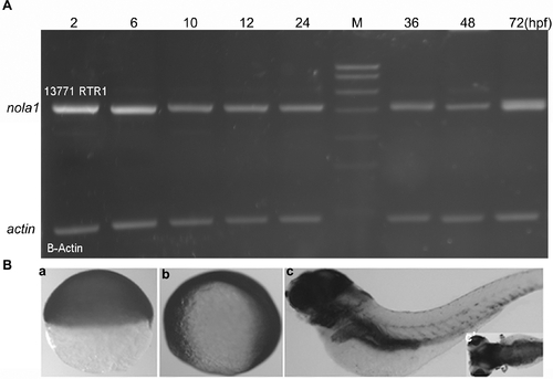
Temporal and spatial expression pattern of nola1. (A) Semi-quantitative PCR result showed that nola1 was expressed from about 2 hpf. (B) Non-specific expression of nola1 was detected during the early stage of zebrafish development (a, b). Afterward, nola1 expression was mainly detected in the brain and some inner organs. a: nola1 expression at 7 hpf; b: nola1 expression at 10 hpf; c and c2: nola1 expression at 4 dpf. a, b: lateral view; c: lateral view with anterior to the left; c2: dorsal view with anterior to the left. |
Expression Data
Expression Detail
Antibody Labeling
Phenotype Data
Phenotype Detail
Acknowledgments
This image is the copyrighted work of the attributed author or publisher, and
ZFIN has permission only to display this image to its users.
Additional permissions should be obtained from the applicable author or publisher of the image.
Full text @ PLoS One