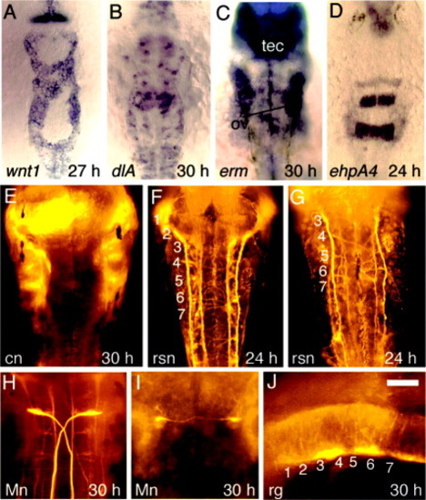
Hindbrain development in tcf3b morphants. All embryos are tcf3b morphants except in H, which depicts a wild-type embryo. A: Expression of wnt1 at 27 hours postfertilization (hpf). B: Expression of dlA at 30 hpf. C: Expression of erm at 30 hpf. The otic vesicles (ov) and tectum (tec) are indicated. D: Expression of ephA4 at 24 hpf. E: Immunostaining with zn8 antibody at 30 hpf shows that commissural neurons (cn) fail to form. Staining of segmental structures along the lateral edges of the hindbrain marks pharyngeal arches and portions of cranial ganglia just out of the plane of focus. F,G: Immunostaining of reticulospinal neurons (rsn) at 24 hpf with anti-acetylated tubulin shows varying degrees of patterning defects in tcf3b-morphants. H,I: 3A10 antibody staining of Mauthner neurons (Mn) at 30 hpf in a wild-type embryo (H) and a tcf3b morphant (I). J: Immunostaining of radial glia (rg) with zrf1-zrf4. Numbers mark locations of the corresponding rhombomeres. Images show dorsal views with anterior to the top (A-I) or a lateral view with anterior to the left (J). Scale bar in J = 60 μm in E,H,I, 75 μm in D,F,G,J, 120 μm in A-C.
|
