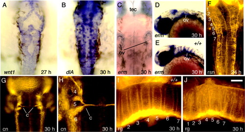
Hindbrain development in spg mutants. All embryos are spg homozygous mutants except for E and I, which show wild-type controls. A: wnt1 expression at 27 hours postfertilization (hpf). B: dlA expression at 30 hpf. C-E: erm expression at 30 hpf in spg mutants (C,D) and a wild-type embryo (E). Spatial patterns are highly variable between spg mutants, but the level of expression is invariably reduced. For landmarks, the otic vesicles (ov) and tectum (tec) are indicated. F: Reticulospinal neurons (rsn) labeled with anti-acetylated tubulin at 24 hpf. G,H: Commissural neurons (cn) labeled with zn8 antibody at 30 hpf. Commissural neurons are absent in anterior rhombomeres and in posterior rhombomeres produce aberrant commissures (c) through the center of the r5 region. H is an enlargement of the left side of the specimen shown in G. I,J: Radial glia (rg) labeled with zrf1-4 antibodies at 30 hpf in a wild-type embryo (I) and a spg mutant (J). The wild-type rg pattern is an enlargement of the specimen shown in Figure 2O. Numbers indicate the positions of corresponding rhombomeres. Images show dorsal views with anterior to the top (A-C,F-H) or a lateral view with anterior to the left (D,E,I,J). Scale bar in J = 25 μm in H, 50 μm in I,J, 65 μm in A-C,F,G, 150 μm in D,E.
|
