
Fig. 4
- ID
- ZDB-FIG-090710-71
- Publication
- Aanstad et al., 2009 - The Extracellular Domain of Smoothened Regulates Ciliary Localization and Is Required for High-Level Hh Signaling
- Other Figures
- All Figure Page
- Back to All Figure Page
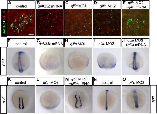
Inhibition of Ciliogenesis Causes Loss of Hh Target Gene Expression in Zebrafish Embryos (A–E) Expression of the ciliary marker acetylated tubulin (green) and the basal body marker γ-tubulin (red) in Kupffer′s vesicles of 16 hpf zebrafish embryos. Injection of dnKif3b mRNA (B), qilin morpholino 1 (MO1) (C), or qilin MO2 (D) caused a reduction of KV cilia, which were restored in embryos coinjected with qilin MO2 and qilin mRNA (E). Scale bar in (A) represents 10 μm. (F–O) In situ hybridization of 10 hpf control and injected embryos. (F–M) In WT embryos, ptc1 (F) and myod (K) are expressed in two stripes flanking the dorsal midline. Injection of dnKif3b (G), qilin MO1 (H), or qilin MO2 (I and L) caused a reduction or loss of ptc1 and myod expression, which could be restored by coinjection of qilin MO2 with qilin mRNA (J and M). (N and O) sonic hedgehog (shh) expression in qilin MO2-injected embryos (O) was comparable to controls (N), with a broadened expression reflecting convergence-extension defects. For quantification of these experiments, see Figure S10. |
| Genes: | |
|---|---|
| Antibody: | |
| Fish: | |
| Knockdown Reagents: | |
| Anatomical Terms: | |
| Stage Range: | Bud to 14-19 somites |
| Fish: | |
|---|---|
| Knockdown Reagents: | |
| Observed In: | |
| Stage Range: | Bud to 14-19 somites |