
Fig. 3
- ID
- ZDB-FIG-090710-70
- Publication
- Aanstad et al., 2009 - The Extracellular Domain of Smoothened Regulates Ciliary Localization and Is Required for High-Level Hh Signaling
- Other Figures
- All Figure Page
- Back to All Figure Page
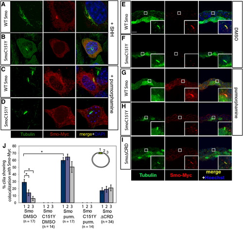
SmoC151Y Does Not Localize to the Cilium Expression of the ciliary marker acetylated tubulin (green) and either Myc-tagged WT Smo or Myc-tagged SmoC151Y (red) in NIH 3T3 cells (A–D) and zebrafish embryos at 10 hpf (E–J). Nuclei of NIH 3T3 cells were visualized with DAPI (blue). (A and B) In NIH 3T3 cells, WT Smo localized to the cilium in response to Hh (A), whereas SmoC151Y did not (B). (C and D) Treatment with the Smo agonist purmorphamine induced ciliary localization of WT Smo (C) but did not induce detectable ciliary localization of SmoC151Y (D). (E–I) Myc-tagged Smo mRNA was injected into Tg(-1.8gsc:GFP)ml1 zebrafish embryos, which express GFP in the dorsal midline. WT Smo localized to the cilia of cells surrounding the dorsal midline in embryos treated with DMSO (E), whereas SmoC151Y was not detected on the cilium (F). Purmorphamine treatment increased the ciliary localization of Smo (G) but did not induce the ciliary localization of SmoC151Y (H). In contrast, SmoΔCRD localized to the cilia in untreated embryos (I). Scale bar in (E) represents 10 μm. (J) The ciliary localization of Smo was quantified in locations away from the dorsal midline in sections (upper right), and the mean percentages of cilia exhibiting colocalization with the Myc-tagged Smo constructs are shown. Error bars indicate SEM. Total numbers of sections counted are shown below. |