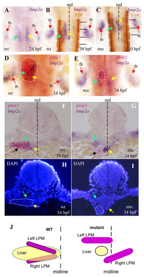
The mypt1sq181 mutation alters the spatial alignment between the liver primordium and the two stripes of LPM expressing Bmp2a. (A-C) Dorsal view of zebrafish embryos subject to WISH using the bmp2a probe in wild type (wt) and mutant (mu). Somites were stained with the F59 antibody (B,C, horizontal black lines). (D,E) Dorsal view of WISH embryos using prox1 (red staining of the liver primordium, circled with a white dashed line) and bmp2a (purple) probes. (F-I) Sectioning of the prox1 and bmp2a WISH embryos at 34 hpf (F,G) and corresponding DAPI staining (H,I). Green and yellow arrows mark the left and right stripes of LPM, respectively; black arrows mark the liver primordium; red arrows mark the fin buds (fb). The white dotted line circles the liver primordium and the black dashed line shows the midline (md). (J) Schematic showing how the liver primordium at 34 hpf is sandwiched between the two bmp2a stripes in wild-type (WT) embryos, one above the primordium and one beneath, whereas in the mypt1sq181 mutant the right stripe fails to cross the midline to align with the left stripe and sandwich the liver primordium.
|
