|
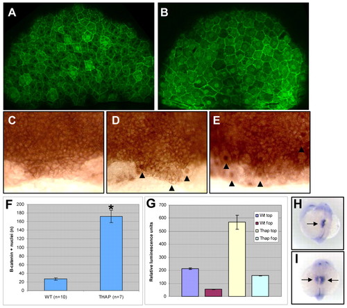
Ca2+ inhibition activates β-catenin and β-catenin is sufficient to alter laterality. Confocal images of β-catenin immunolocalization at 70-80% epiboly in (A) wt and (B) thapsigargin-treated embryos. HRP staining of β-catenin protein oriented on the DFC region in (C) wt, (D) thapsigargin-treated and (E) DFC-targeted Axin-1 MO-injected embryos; arrowheads indicate β-catenin-positive nuclei. (F) Comparison of total nuclear β-catenin in wt and thapsigargin-treated embryos.* indicates P-value of 2.5x10⁻⁵. (G) TopFlash (top) vs. FopFlash (fop) luciferase reporter constructs analyzed for relative luminescence in wt or thapsigargin-treated embryos, normalized to control (Renilla) luciferase. Data are means ± s.e. (H,I) Combined lefty1 and lefty2 expression (arrows) in the left LPM and brain in wt (H) and bilateral (I) in DFC-targeted β-catenin RNA-injected embryos: dorsal-anterior view, 19-23 somites.
|