
Fig. 6
- ID
- ZDB-FIG-080114-24
- Publication
- Schneider et al., 2008 - Calcium fluxes in dorsal forerunner cells antagonize {beta}-catenin and alter left-right patterning
- Other Figures
- All Figure Page
- Back to All Figure Page
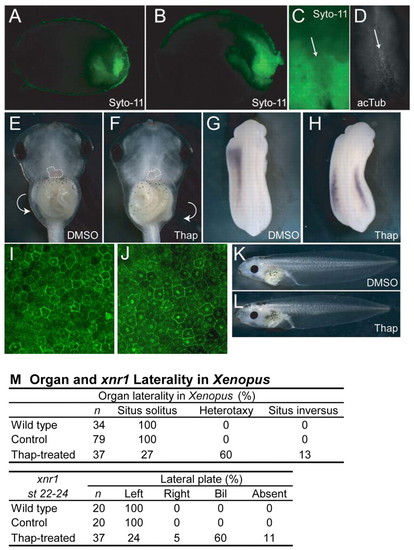
Xenopus DFC-like cells and Ca2+ requirement for laterality. Xenopus embryos incubated with Syto-11 at stage 11.5 and sagittally bisected at (A) stage 14 and (B) stage 17 showing Syto-11-labeled cells enriched in the future tail region. (C) Stage 17 archenteron roof explant, showing Syto-11 staining of the node (arrow) and surrounding LPM. (D) Explant in C immunostained for acetylated tubulin (acTub), which is enriched in the node region (arrow). (E) Ventral view of stage 46 control-treated embryo, showing situs solitus, or normal lateral asymmetry. The heart is outlined, showing the left-sided position of the ventricle and right-sided outflow tract. The gut coils counterclockwise, as shown by the arrow. (F) Ventral view of a thapsigargin-treated embryo at stage 46, showing an example of situs inversus, or reversed laterality. The heart is outlined, showing the right-sided position of the ventricle and left-sided outflow tract. The gut coils clockwise, as shown by the arrow. (G) Left-sided xnr1 expression at stage 22-24 in a DMSO-treated embryo. (H) Bilateral xnr1 expression in a thapsigargin-treated embryo. (I,J) Lateral view of β-catenin protein localization in control-treated (I) and thapsigargin-treated (J) stage 13 embryos. Note increased number and intensity of nuclear staining. (K,L) Lateral views of control (K) and thapsigargin-treated (L) embryos, showing normal axial development. (M) Summary of Xenopus laterality data. Thap, thapsigargin. |