
Fig. 3
- ID
- ZDB-FIG-071228-19
- Publication
- Veldman et al., 2007 - Gene expression analysis of zebrafish retinal ganglion cells during optic nerve regeneration identifies KLF6a and KLF7a as important regulators of axon regeneration
- Other Figures
- All Figure Page
- Back to All Figure Page
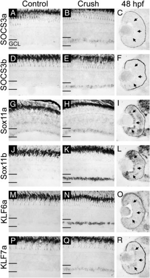
In situ hybridization verification of microarray results and comparison with expression in the developing retina (part II). mRNA for SOCS3a (A–C), SOCS3b (D–F), Sox11a (G–I), Sox11b (J–L), KLF6a (M–O), and KLF7a (P–R) was detected by in situ hybridization in uninjured control, 3-day post-optic nerve crush and 48 hpf retinal sections. Expression of all six genes is elevated in the ganglion cell layer (GCL) following optic nerve crush injury as compared to control. Developmentally, Sox11a, Sox11b, and KLF7a are expressed in the GCL at 48 hpf while SOCS3a, SOCS3b, and KLF6a are not (arrows in panels C, F, I, L, O, and R). Sense probes for all genes tested gave no specific signal (data not shown). |
| Genes: | |
|---|---|
| Fish: | |
| Condition: | |
| Anatomical Term: | |
| Stage Range: | Long-pec to Adult |
Reprinted from Developmental Biology, 312(2), Veldman, M.B., Bemben, M.A., Thompson, R.C., and Goldman, D., Gene expression analysis of zebrafish retinal ganglion cells during optic nerve regeneration identifies KLF6a and KLF7a as important regulators of axon regeneration, 596-612, Copyright (2007) with permission from Elsevier. Full text @ Dev. Biol.