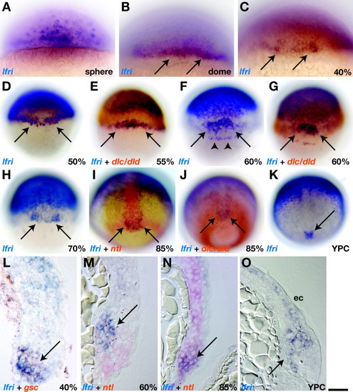
Dorsal margin cells transiently express lfng during gastrulation. A,B: Lateral views; C-K: dorsal views; and L-O: sagittal sections of blastula and gastrula stage embryos processed for in situ RNA hybridization. A: Sphere stage (4 hours postfertilization [hpf]). lfng expression was punctate throughout the blastoderm. B: Dome stage (4.3 hpf). Arrows indicate lfng+ cells near the embryonic margin. C,D: Arrows indicate lfng+ cells concentrated at the dorsal margin at 40% epiboly (5 hpf) and 50% epiboly (5.3 hpf). E: At 55% epiboly, lfng+ cells occupied a more narrow area of the dorsal margin (arrows) and appeared to be adjacent to cells that expressed dlc and dld (red). F: At 60% epiboly (6 hpf). Arrows indicate lfng+ cells located just above the dorsal margin, and arrowheads mark lfng+ dorsal forerunner cells. G: Dorsal lfng+ cells appeared to be adjacent to dlc- and dld-expressing cells. H: At 70% epiboly (8 hpf). Arrows mark lfng+ cells at the dorsal margin. I,J: At 85% epiboly (8.6 hpf). Embryos continued to express lfng at the dorsal margin (arrows), in cells that also expressed ntl (I) and that were adjacent to dlc/dld+ cells (J). K: At yolk plug closure (YPC) stage (10 hpf). Arrow indicates lfng expression at dorsal margin. L: At 40% epiboly. Dorsal margin cells (arrow) coexpressed gsc (red) and lfng (blue). M: At 60% epiboly lfng+ cells (blue, arrow) occupied dorsal mesendoderm, just anterior to ntl+ cells (red). N: At 85% epiboly. Dorsal margin cells (arrow) coexpressed lfng (blue) and ntl (red). O: YPC stage. lfng+ cells were deep in the nascent tail bud (arrow) and in ectoderm (ec). Scale bar = 80 μm in A-C, 160 μm in D-K, 40 μm in L-O.
|
