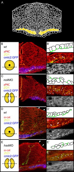
Apicobasal polarity is disrupted in has/prkci and nok/mpp5 morphants. Transverse sections of heart cone stage (20-somites) embryos. The section plane is indicated in each case (dotted lines). PRKC/aPKC and <α-catenin, red or gray; ZO-1, green; GFP false colored in blue. (A) Drawing of a transverse section through the center of the heart cone. Two bilateral wings of myocardial cells are indicated in yellow. (B,B'') In wild type, ZO-1 is localized to the apical membranes of the monolayered myocardial cell layer. (B''') PRKC is localized basal of ZO-1 (see arrowheads). (C,C'') nok morphants display diffusely positioned spots of ZO-1. (C''') PRKC is mislocalized in a diffuse pattern. (D,D'',D''') Co-localization of ZO-1 and α-catenin at the apical membrane in wild type. (E,E'',E''') In has morphants, co-localization of ZO-1 and ß-catenin is lost. The peridermal localization of ZO-1 and α-catenin is not affected (arrowheads). (E''') α-catenin is diffusely distributed along the membrane. Stars indicate myocardial cells.
|
