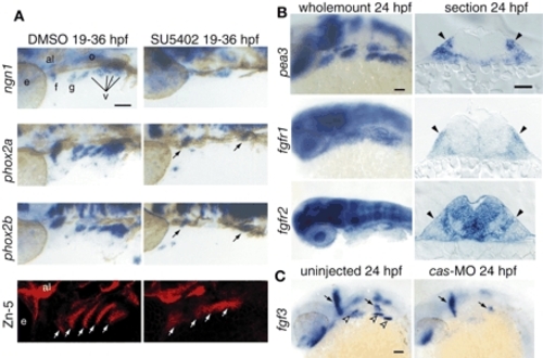
Fgf signaling is active in EB placodes. (A) Zebrafish embryos treated with the Fgfr inhibitor SU5402 (right) or DMSO (left) beginning at 19 hpf were collected at 36 hpf and processed for in-situ hybridization with ngn1, phox2a and phox2b. All panels show lateral views. Anterior is at right. In the SU5402-treated embryos, ngn1, phox2a and phox2b expression is either reduced (arrows) or absent. Fgfr inhibitor treatment also affected pharyngeal pouch morphology and size, as assessed by Zn-5 immunolabeling (white arrows). (B) Expression analyses of pea3, fgfr1 and fgfr2 at 24 hpf. Whole-mount panels are lateral views of the embryos; transverse sections are at the level of the facial placode. Note characteristic ectodermal thickenings, indicating cranial placodes (black arrowheads). pea3 and fgfr1 were expressed in the EB placodes, while fgfr2 was not. (C) fgf3 was expressed in the pharyngeal endoderm (left; empty arrowheads) at 24 hpf. As expected, the endodermal fgf3 expression was lost in cas morphants (right). Scale bars: 50 µm. Abbreviations are as in Fig. 1.
|
