
- Title
-
Nonmuscle myosin II-B (myh10) expression analysis during zebrafish embryonic development
- Authors
- Huang, Y., Wang, X., Wang, X., Xu, M., Liu, M., and Liu, D.
- Source
- Full text @ Gene Expr. Patterns
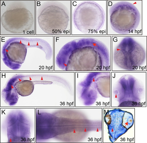
Whole mount in situ hybridization analysis of zebrafish embryos (1 cell–36 hpf) using antisense Danio rerio myh10 probe. (A) 1 cell, lateral view, no hybridization signal. (B) 50% epiboly, lateral view, no hybridization signal. (C) 75% epiboly, lateral view, no hybridization signal. (D) 14 hpf, lateral view, hind brain (arrowhead). (E) 20 hpf, lateral view, central nervous system (arrowheads). (F) 20 hpf, lateral view, eye (arrowhead), rhombomere (arrow). (G) 20 hpf, dorsal view, midbrain (arrowhead). (H) 36 hpf, lateral view, brain and spinal cord (arrowheads). (I) 36 hpf, lateral view, otic vesicle (arrow head). (J) 36 hpf, ventral view, eye (arrowhead). (K) 36 hpf, dorsal view, otic vesicle (arrow head). (L) 36 hpf, dorsal view, spinal cord (arrowheads). (M) Section of 36 hpf head, retina (arrow). |
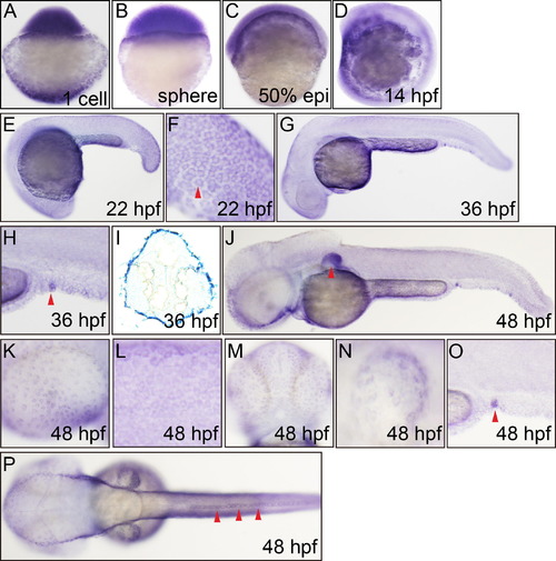
Whole mount in situ hybridization analysis of zebrafish embryos (1 cell-48 hpf) using antisense Danio rerio myh9a probe. (A) 1 cell, lateral view, strong staining. (B) Sphere, lateral view. (C) 50% epiboly, lateral view. (D) 14 hpf, lateral view, epidermis. (E) 22 hpf, lateral view, epidermis, enveloping layer (EVL). (F) 22 hpf, lateral view, caudal trunk, EVL (arrowhead). (G) 36 hpf, lateral view, epidermis of the whole body. (H) 36 hpf, lateral view, urogenital opening (arrowhead). (I) 36 hpf, transverse section of head, out layer staining. (J) 48 hpf, lateral view, pectoral fin (arrowhead). (K) 48 hpf, lateral view, eye epidermis. (L) 48 hpf, lateral view, trunk epidermis over the yolk tube elongation. (M) 48 hpf, ventral view, head epidermis. (N) 48 hpf, lateral view, pectoral fin epidermis. (O) 48 hpf, lateral view, urogenital opening (arrowhead). (P) 48 hpf, dorsal view, dorsal fin epidermis (arrowheads). |

Expression pattern of myh10 was analyzed by whole mount in situ hybridization and whole mount antibody staining at 48 hpf and 72 hpf stages embryos. (A) 48 hpf, lateral view, myh10 expression in brain and anterior spinal cord. (B) 48 hpf, lateral view, retina (arrow), ear (arrowhead). (C) 48 hpf, ventral view, retina (arrow) and olfactory bulb (arrowhead). (D) 48 hpf, dorsal view, ear (arrowhead). (E) 48 hpf, lateral view, heart (arrowhead). (F) 48 hpf, lateral view, retina (arrow) and olfactory bulb (arrowhead). (G) 72 hpf, lateral view. (H) 72 hpf, lateral view, retina (arrowhead). (I) 72 hpf, ventral view, retina (arrowhead), olfactory bulb neurons (arrow). (J) 72 hpf, lateral view, otic capsule. EXPRESSION / LABELING:
|
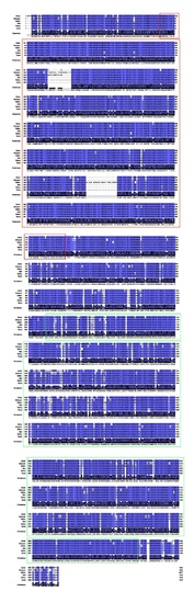
Myosin_head (motor domain), and Myosin_tail_1 of NM II-B are highly conserved during vertebrates. Alignment of Myosin N-terminal SH3-like domain amino acid residue sequences of Danio rerio, Xenopus laevis, Gallus gallus, Mus musculus, Rattus norvegicus and Homo sapiens NM II-B. The sequences are retrieved from NCBI Protein sequence database. The sequences accessions IDs are listed following respectively, XP_683046.5, NP_001084034.1, NP_990805.1, NP_780469.1, NP_113708.1, NP_005955.3. These protein sequences were aligned using T-COFFEE and edited by Jalview. Myosin_head domain sequence is indicated in red rectangle. Myosin_tail_ is in green rectangle. |

Whole mount in situ hybridization analysis of zebrafish embryos (2 cells-48 hpf) using antisense Danio rerio myh9b probe. A. 2 cells, strong staining. B. 4 cells, strong staining. C. 1K cells, strong staining. D. 50% epiboly, lateral view. E. 14 hpf, lateral view, epidermis. F. 24 hpf, lateral view, epidermis, enveloping layer (EVL). G. 24 hpf, lateral view, caudal trunk, EVL (arrowhead). H. 36 hpf, lateral view, epidermis of the whole body. I. 36 hpf, lateral view, head epidermis. J. 36 hpf, lateral view, trunk epidermis. K. 48 hpf, lateral view, epidermis of the whole body, pectoral fin epidermis (arrowhead). L. 48 hpf, dorsal view, pectoral fin epidermis (arrowhead). |
Reprinted from Gene expression patterns : GEP, 13(7), Huang, Y., Wang, X., Wang, X., Xu, M., Liu, M., and Liu, D., Nonmuscle myosin II-B (myh10) expression analysis during zebrafish embryonic development, 265-270, Copyright (2013) with permission from Elsevier. Full text @ Gene Expr. Patterns