
- Title
-
Functional Analysis of Retinitis Pigmentosa 2 (RP2) Protein Reveals Variable Pathogenic Potential of Disease-Associated Missense Variants
- Authors
- Patil, S.B., Hurd, T.W., Ghosh, A.K., Murga-Zamalloa, C.A., and Khanna, H.
- Source
- Full text @ PLoS One
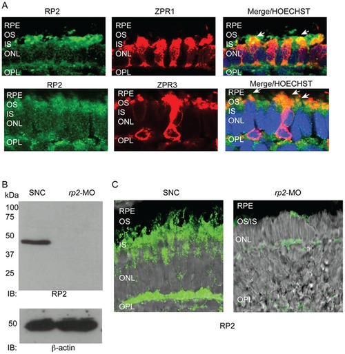
RP2 is expressed in zebrafish photoreceptors. A. Retinal cryosections from 4 dpf zebrafish embryos were analyzed by immunofluorescence using anti-Rp2 antibody (green). Photoreceptor markers, Zpr1 and Zpr3 (red) were used to stain cone and rod photoreceptors, respectively. Arrows in Merge indicate co-localization (yellow) of RP2 with both markers. Nuclei are marked with Hoechst (blue). RPE: retinal pigmented epithelium; OS: outer segment; IS: inner segment; ONL: outer nuclear layer; OPL: outer plexiform layer. B. Protein extracts from 4 dpf zebrafish embryos injected with standard negative control (SNC) morpholino (MO) or rp2-MO were analyzed by SDS-PAGE and immunoblotting using anti-RP2 antibody. The expected size band of 42 kDa was detected in the SNC-treated embryo extracts but not in the rp2-MO treated ones. Lower panel shows immunoblotting of same samples using anti-β actin antibody, as loading control. C. Immunofluorescence analysis of cryosections of SNC and rp2-mo treated embryo retina (4 dpf) was performed using anti-RP2 antibody (green). Merge with Nomarski image shows that the expression of RP2 was significantly down regulated in the rp2-MO treated embryos. EXPRESSION / LABELING:
|
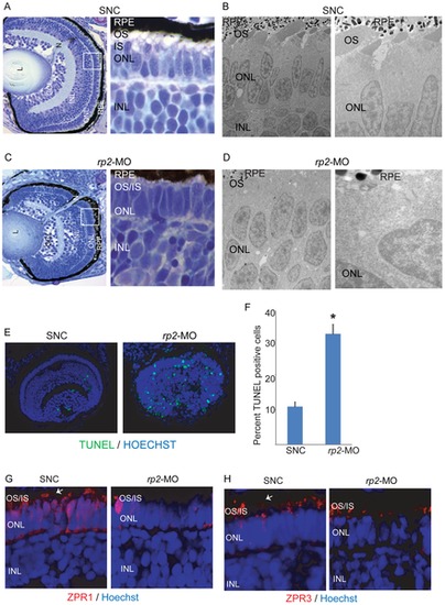
Silencing of rp2 results in retinopathies in zebrafish. A & C. Histological analysis of zebrafish retina treated with SNC (A) or rp2-MO (D) was performed at 4 dpf. Insets show enlarged image of the area marked in white square. B & D. Electron microscopy analysis of SNC (B) and rp2-MO (D) treated embryo retinas shows defective OS development due to knockdown of rp2. Right panels are higher magnification images of the left panels. E. TUNEL staining of the indicated zebrafish retinas was performed using manufacturer′s instructions. Green nuclei indicate pycknotic cells. The TUNEL staining was quantified and represented in panel F. *: p<0.001. G, H. SNC and rp2-MO treated embryo retina cryosections were stained with Zpr1 and Zpr3 antibodies (red; arrows). Nuclei were stained with Hoechst (blue). OS: outer segment; IS: inner segment; ONL: outer nuclear layer; INL: inner nuclear layer. PHENOTYPE:
|

Human WT RP2 can rescue rp2-MO associated phenotype. A. Histological analysis of retinas from embryos co-injected with rp2-MO and mRNA encoding GFP (left panel; rp2-MO + GFP) or wild type (WT) RP2-GFP fusion protein (right panel; rp2-MO + RP2-GFP). OS: outer segment; IS: inner segment; ONL: outer nuclear layer; OPL: outer plexiform layer. B and C. Immunofluorescence analysis of the indicated embryo retinas was performed using Zpr1 or Zpr3 antibodies (red; arrows) or anti-GFP antibodies (green). Hoechst was used to stain nuclei (blue). OS: outer segment; IS: inner segment; ONL: outer nuclear layer; INL: inner nuclear layer; GCL: ganglion cell layer. |
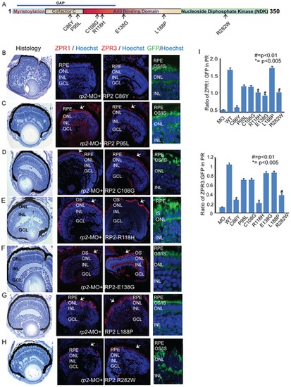
RP2 disease mutations exhibit diverse pathogenic potential. A. Schematic representation of the primary structure of the RP2 protein and predicted domains. Arrows indicate the mutations tested in this study. GAP: GTPase activating protein domain. B–H. Histological and immunofluorescence analyses (using ZPR1 or ZPR3 antibodies; red; arrows or anti-GFP; green) of retinas of embryos injected with rp2-MO and mRNA encoding indicated RP2 mutant proteins (fused to GFP) was performed as described in the Methods section. Nuclei are stained with Hoechst (blue). RPE: retinal pigmented epithelium; OS: outer segment; IS: inner segment; ONL: outer nuclear layer; INL: inner nuclear layer; GCL: ganglion cell layer. I. Quantitative analysis of ZPR1 and ZPR3 expression in the photoreceptor layer of the different embryos was performed and compared to expression of the WT or mutant RP2-GFP protein. The results are represented as ratio of Zpr1 or Zpr3 expression to the GFP expression in photoreceptors. |

RP2 is expressed in both rod and cone photoreceptors. Immunofluorescence analysis of 4 dpf zebrafish embryos injected SNC-MO and rp2-MO was performed using anti-rhodopsin (1D4) antibody or PNA (red). Nuclei are stained with DAPI (blue). RPE: retinal pigmented epithelium; OS: outer segment; IS: inner segment; ONL: outer nuclear layer; INL: inner nuclear layer; GCL: ganglion cell layer. PHENOTYPE:
|