
|
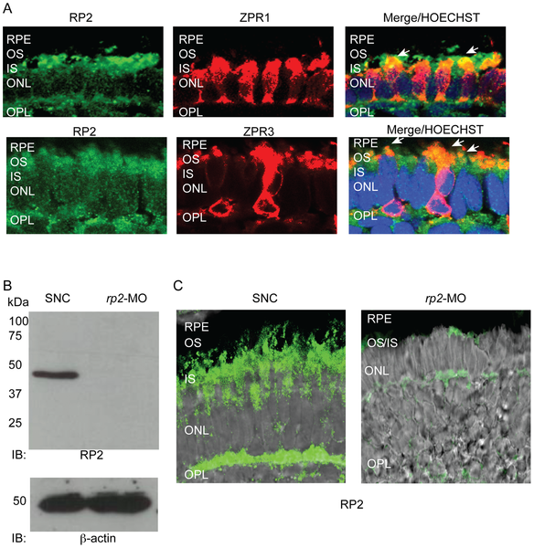
Fig. 1
RP2 is expressed in zebrafish photoreceptors.
A. Retinal cryosections from 4 dpf zebrafish embryos were analyzed by immunofluorescence using anti-Rp2 antibody (green). Photoreceptor markers, Zpr1 and Zpr3 (red) were used to stain cone and rod photoreceptors, respectively. Arrows in Merge indicate co-localization (yellow) of RP2 with both markers. Nuclei are marked with Hoechst (blue). RPE: retinal pigmented epithelium; OS: outer segment; IS: inner segment; ONL: outer nuclear layer; OPL: outer plexiform layer. B. Protein extracts from 4 dpf zebrafish embryos injected with standard negative control (SNC) morpholino (MO) or rp2-MO were analyzed by SDS-PAGE and immunoblotting using anti-RP2 antibody. The expected size band of 42 kDa was detected in the SNC-treated embryo extracts but not in the rp2-MO treated ones. Lower panel shows immunoblotting of same samples using anti-β actin antibody, as loading control. C. Immunofluorescence analysis of cryosections of SNC and rp2-mo treated embryo retina (4 dpf) was performed using anti-RP2 antibody (green). Merge with Nomarski image shows that the expression of RP2 was significantly down regulated in the rp2-MO treated embryos.