
Fig. 2
- ID
- ZDB-FIG-110720-45
- Publication
- Patil et al., 2011 - Functional Analysis of Retinitis Pigmentosa 2 (RP2) Protein Reveals Variable Pathogenic Potential of Disease-Associated Missense Variants
- Other Figures
- All Figure Page
- Back to All Figure Page
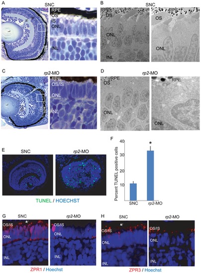
Silencing of rp2 results in retinopathies in zebrafish. A & C. Histological analysis of zebrafish retina treated with SNC (A) or rp2-MO (D) was performed at 4 dpf. Insets show enlarged image of the area marked in white square. B & D. Electron microscopy analysis of SNC (B) and rp2-MO (D) treated embryo retinas shows defective OS development due to knockdown of rp2. Right panels are higher magnification images of the left panels. E. TUNEL staining of the indicated zebrafish retinas was performed using manufacturer′s instructions. Green nuclei indicate pycknotic cells. The TUNEL staining was quantified and represented in panel F. *: p<0.001. G, H. SNC and rp2-MO treated embryo retina cryosections were stained with Zpr1 and Zpr3 antibodies (red; arrows). Nuclei were stained with Hoechst (blue). OS: outer segment; IS: inner segment; ONL: outer nuclear layer; INL: inner nuclear layer. |
| Fish: | |
|---|---|
| Knockdown Reagent: | |
| Observed In: | |
| Stage: | Day 4 |