
Fig. 4
- ID
- ZDB-FIG-110720-47
- Publication
- Patil et al., 2011 - Functional Analysis of Retinitis Pigmentosa 2 (RP2) Protein Reveals Variable Pathogenic Potential of Disease-Associated Missense Variants
- Other Figures
- All Figure Page
- Back to All Figure Page
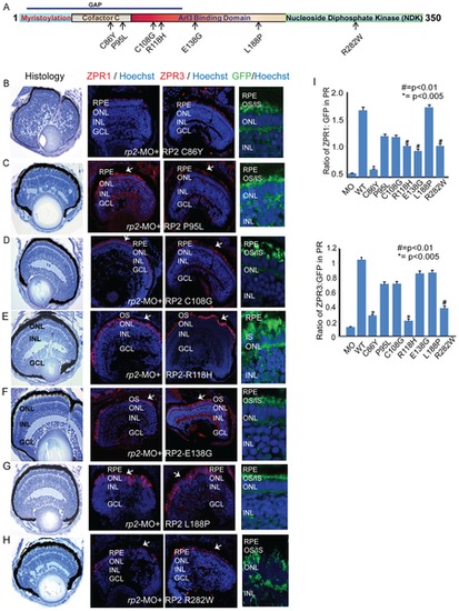
RP2 disease mutations exhibit diverse pathogenic potential. A. Schematic representation of the primary structure of the RP2 protein and predicted domains. Arrows indicate the mutations tested in this study. GAP: GTPase activating protein domain. B–H. Histological and immunofluorescence analyses (using ZPR1 or ZPR3 antibodies; red; arrows or anti-GFP; green) of retinas of embryos injected with rp2-MO and mRNA encoding indicated RP2 mutant proteins (fused to GFP) was performed as described in the Methods section. Nuclei are stained with Hoechst (blue). RPE: retinal pigmented epithelium; OS: outer segment; IS: inner segment; ONL: outer nuclear layer; INL: inner nuclear layer; GCL: ganglion cell layer. I. Quantitative analysis of ZPR1 and ZPR3 expression in the photoreceptor layer of the different embryos was performed and compared to expression of the WT or mutant RP2-GFP protein. The results are represented as ratio of Zpr1 or Zpr3 expression to the GFP expression in photoreceptors. |