
- Title
-
Essential Role for the d-Asb11 cul5 Box Domain for Proper Notch Signaling and Neural Cell Fate Decisions In Vivo
- Authors
- Sartori da Silva, M.A., Tee, J.M., Paridaen, J., Brouwers, A., Runtuwene, V., Zivkovic, D., Diks, S.H., Guardavaccaro, D., and Peppelenbosch, M.P.
- Source
- Full text @ PLoS One

Phenotypic assays on wild type and asb11cul embryos. (A), Morphological analysis of wild type and mutant embryos at 48 and 72hpf. (B), (left) Anterior view of wild type and mutant embryos at 10hpf after whole mount in situ hybridization, WISH, using probe against d-asb11. (right) Graph shows the quantification of the respective expressions using qPCR. (C), (left) Endogenous d-Asb11 in wild type (WT), heterozygous (asb11+/-) and mutant (asb11cul) embryos at 12 hpf was detected by immunoblotting using anti-d-Asb11 antibody. (right) Graph quantifies 3 individual experiments, with 30 embryos/genotype/experiment. EXPRESSION / LABELING:
PHENOTYPE:
|
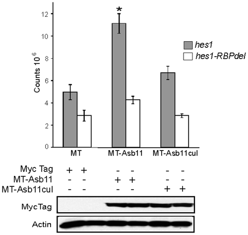
Cullin box domain promotes induction of hes1 gene in vitro. nTera-d1 cells were co-transfected with hes1-luciferase (hes1) or hes1-luciferase lacking the conserved CSL-binding site (hes1-RBPdel) and myc-tag (MT) as a control, or myc-tagged d-asb11 full length (MT-Asb11) or myc-tagged asb11cul (MT-Asb11cul) cDNA. Hes1-dependent Notch activity was analyzed by luciferase measurement. |
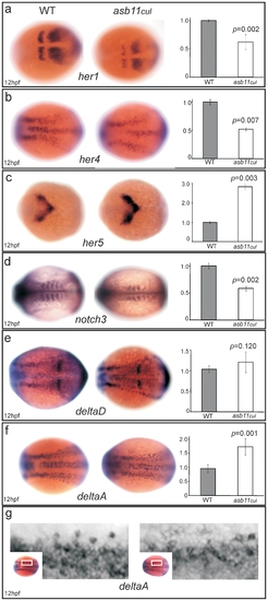
asb11cul presented altered expression of Delta-Notch pathway components. Wild type (left panel) and mutant (middle panel) embryos at 12 hpf were analyzed for WISH using probes against her1, A; her4, B; her5, C; notch3, D; deltaD, E; and deltaA, F. (G), Higher magnification shows detailed analysis of deltaA expression. (left) Graphs quantify the mRNA expression levels. EXPRESSION / LABELING:
|
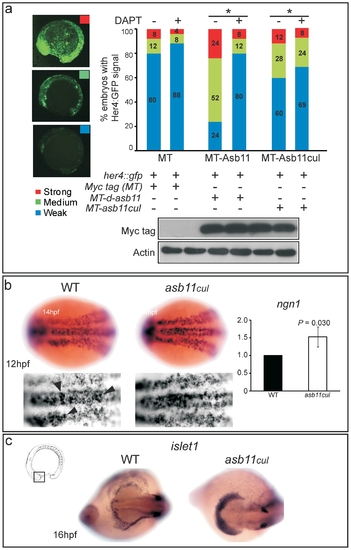
her4::gfp transactivation and premature differentiation of neural cells in asb11cul. (A), the her4::gfp reporter was co-injected with myc-tag (MT) mRNA as a control, myc-tagged d-asb11 full length (MT-Asb11) or myc-tagged asb11cul (MT-Asb11cul) mRNA in zebrafish embryos. Injected embryos were treated with (+) (n = 25) or without (-) (n = 25) DAPT, from 1.5 hpf. At 14 hpf, embryos were analyzed for her4 transactivation based on the intensity of the GFP signal. Positive embryos were counted and percentages of embryos presenting weak (blue), medium (green) or strong (red) signal were given. (B), Wild type (left panel) and mutant (middle panel) embryos at 12 hpf were analyzed for WISH using probe against ngn1. (right) Graph quantifies expression of ngn1 using qPCR. (C) Wild type (left panel) and mutant (right panel) polster of embryos at 16 hpf were analyzed for WISH using probe against islet1. EXPRESSION / LABELING:
|
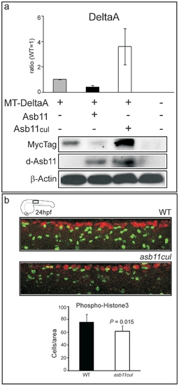
Cullin box is essential for DeltaA degradation and for maintaining a cell proliferating state in vivo. (A) Zebrafish embryos were injected with Myc-tagged deltaA (MT-DeltaA) and d-asb11 (Asb11) or asb11cul (Asb11cul) mRNA at one-cell stage. (lower panel) Lysates of 12 hpf embryos were analyzed by immunoblotting for the presence of DeltaA. (higher panel) Graph quantifies 2 individual experiments, each with 30 injected embryos/group. (B), Fluorescent whole-mount antibody labeling of wild type (WT) and asb11cul embryos at 24 hpf for the mitotic marker anti-phosphohistone-3 (PH 3) antibody (green) and the neuronal marker Hu(C). Graph shows the number of positive cells per area (5 somites from beginning of yolk extension) of 5 embryos for each genotype. |