
- Title
-
The activation of membrane targeted CaMK-II in the zebrafish Kupffer's vesicle is required for left-right asymmetry
- Authors
- Francescatto, L., Rothschild, S.C., Myers, A.L., and Tombes, R.M.
- Source
- Full text @ Development
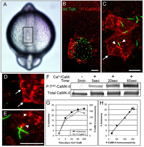
CaMK-II is activated on the left side of Kupffer′s vesicle (KV). (A) Dorsal view of a 12-somite embryo, shows the KV (at the bottom of outlined rectangle) by differential interference contrast microscopy. (B) Confocal immunofluorescence of rectangular region in A using anti-acetylated α-tubulin (green, Alexa 488) to localize cilia and anti-P-T287 CaMK-II (red, Alexa 568) to localize activated CaMK-II in cells on the left side of the KV. (C-E) Higher magnification reveals activated CaMK-II along the cell cortex (arrows) and intracellular clusters (arrowheads), which occasionally colocalize with the base of cilia. Scale bars 10 μm. (F-H) The anti-P-T287 antibody reacts only with activated CaMK-II, as demonstrated by incubating ectopically expressed zebrafish β1K CaMK-II with Ca2+/CaM for the indicated times and then assessing (F) immunoreactivity with anti-P-T287 CaMK-II and an antibody reactive with total CaMK-II. (G) CaMK-II autonomy, measured by peptide assay, and P-T287 immunoreactivity for a representative experiment. (H) When values from four experimental replicates were compiled and plotted against each other, P-T287 CaMK-II immunoreactivity (blot density) was proportional to autonomy. EXPRESSION / LABELING:
|

Left-sided CaMK-II activation is transient. (A-E) The top row shows representative confocal immunofluorescent projections of P-T287 CaMK-II (red) and cilia (green) at the three- (A), six- (B), 10- (C), 14- (D) and 18- (E) somite stages. These projections of z-stacks were conducted as in Fig. 1 (12 somites) and are displayed at the same intensity. Scale bar: 10 μm. Middle row shows KV morphology (asterisk) from the same dorsal perspective. Scale bar: 100 μm. Bottom row shows the position of the KV (arrowheads) from the lateral perspective. (F) For each stage (n=14-36 embryos per stage), the percentage of embryos and the number of cells per embryo that exhibited activated CaMK-II was determined by inspecting z-stacks. (G) The location of KV cells exhibiting activated CaMK-II was scored in one of the four quadrants shown here and results can be found in Tables 1 and 2. EXPRESSION / LABELING:
|
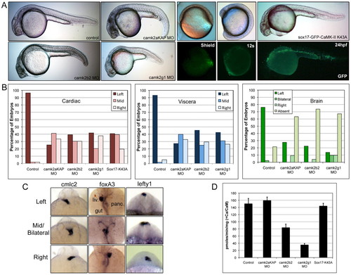
Left-right asymmetry is lost in CaMK-II morphants. (A) Lateral images of representative morphant embryos at 24 hpf and of Sox17-K43A GFP CaMK-II cDNA-injected embryos at shield stage, 12 somites and 24 hpf include fluorescent (GFP) images. (B) Organ asymmetry was scored for embryos injected with 5 ng mismatch, 1.5 ng camk2aKAP, 1.5 ng camk2b2, 1.25 ng camk2g1 MOs and 150 pg Sox17-GFP-CaMK-II K43A cDNA; n=87-233 per condition. (C) Representative dorsal views of camk2g1 morphant embryos stained with probes for cmlc2 (cardiac, 24 hpf), foxa3 (visceral organ, 48 hpf) and lefty1 (epithalamus, <24 somites). (D) Ca2+/CaM-dependent CaMK-II activity in pmoles/minute/mg were determined by peptide assays on 24 hpf lysates after injection with the indicated MO or cDNA. EXPRESSION / LABELING:
PHENOTYPE:
|

CaMK-II mRNAs are uniformly detected around the KV. (A) αKAP encodes an alternative membrane-targeting domain. (B) Mouse and zebrafish αKAP sequences. (C) Dorsal view of camk2aKAP, camk2b2 and camk2g1 in situ hybridization at 12 somites. Asterisk indicates KV location. EXPRESSION / LABELING:
|
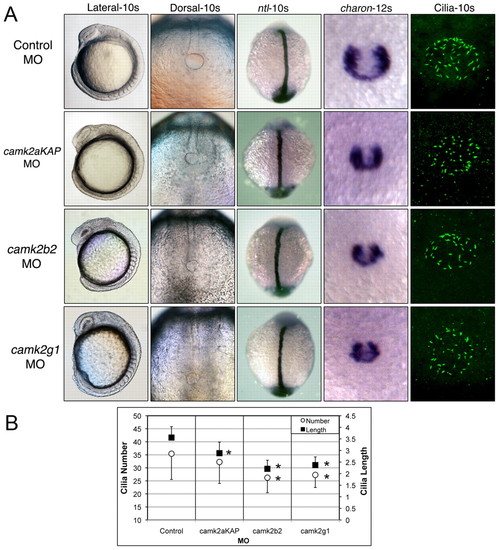
KV markers and cilia in CaMK-II morphants. (A) Embryos were probed with ntl (10s), charon (12s) and anti-acetylated tubulin (Cilia-10s) after injection with 5 ng mismatch (control), 1.5 ng camk2aKAP, 1.5 ng camk2b2 or 1.25 ng camk2g1 MO. (B) Cilia number and length were averaged from 105-201 embryos per condition; *P<0.005 compared with control. EXPRESSION / LABELING:
PHENOTYPE:
|
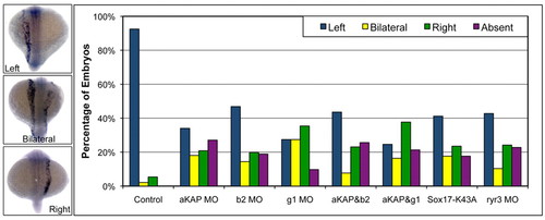
Spaw asymmetry is lost. Left, bilateral, right or absent spaw expression was examined by in situ hybridization (as shown in samples) in single and double morphants of camk2aKAP, camk2b2, camk2g1, ryr3 and sox17-K43A CaMK-II at 18-20 somites and is listed as the percentage of the total embryos in each condition; n=39-145. Control embryos include mismatch MO-injected and buffer-injected embryos. EXPRESSION / LABELING:
PHENOTYPE:
|

CaMK-II activation is disrupted in morphants. Confocal immunofluorescent projections of P-T287 CaMK-II (red) and cilia (green) in embryos at the 12-somite stage after injection with (A) 5 ng mismatch (control), (B) 1.5 ng camk2aKAP, (C) 1.5 ng camk2b2, (D) 1.25 ng camk2g1 MO, (E) 150 ng Sox17-K43A GFP-CaMK-II cDNA (inset is green fluorescence of cilia and GFP-CaMK-II around the KV), (F) 4 ng ntl MO, (G) 4 ng pkd2 MO or (H) 4 ng ryr3 MO. Scale bar: 10 μm. EXPRESSION / LABELING:
PHENOTYPE:
|

Unillustrated author statements PHENOTYPE:
|