
- Title
-
Interaction between thymidylate synthase and its cognate mRNA in zebrafish embryos
- Authors
- Zhang, Y., Yang, S., Liu, M., Song, C., Wu, N., Ling, P., Chu, E., and Lin, X.
- Source
- Full text @ PLoS One
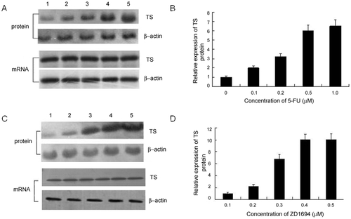
Expression profile of TS mRNA and protein in zebrafish embryos treated with 5-FU or ZD1694. Zebrafish embryos were treated with various concentrations of 5-FU or ZD1694 for 24 h and the expression levels of TS mRNA and protein were evaluated. The TS mRNA and protein level after 5-FU (A, B) or ZD1694 (C, D) treatment is shown. A, Zebrafish embryos were untreated (lane 1) or exposed to 0.1 μM (lane 2), 0.2 μM (lane 3), 0.5 μM (lane 4) or 1.0 μM 5-FU (lane 5) for 24 h, and TS mRNA and protein expression was determined using Northern and Western blotting analysis respectively. C, TS mRNA and protein level in untreated embryos (lane 1) or embryos treated with 0.1 μM (lane 2), 0.2 μM (lane 3), 0.3 μM (lane 4) and 0.4 μM (lane 5) ZD1694, respectively. B and D, The quantification results of protein expression, which represent the protein expression level after 5-FU or ZD1694 treatment, respectively, is shown. The TS expression level in untreated embryos was defined as 1.0. |
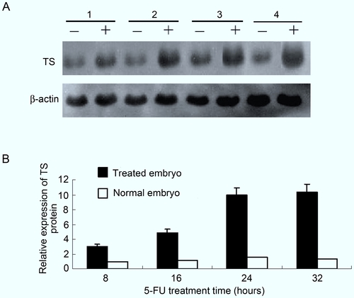
Time-dependent effect of 5-FU on TS expression. Zebrafish embryos (n = 100) were untreated or treated with varying concentrations of 5-FU. A. Western immunoblot analyses were performed to evaluate TS levels in untreated zebrafish embryos or in embryos treated with 1.0 μM 5-FU for 8 h (lane 1), 16 h (lane 2), 24 h (lane 3) and 32 h (lane 4). β-actin was used as internal control. B. The quantification results of protein expression. The expression level of TS in untreated embryos at 8 h post-fertilization (hpf) was defined as 1.0. Each experiment was performed more than three times. |
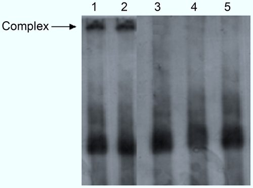
Specific interaction between zebrafish TS and its cognate mRNA in vitro. A gel shift experiment was performed using 32P-labeled full-length TS mRNA as the probe. Zebrafish TS protein (100 ng) was incubated with 32P-labeled full-length TS mRNA (lane 1, arrow). One hundred-fold molar excess luciferase mRNA did not affect the interaction between TS protein and mRNA (lane 2). In lane 3, 100-fold molar excess of unlabeled TS mRNA was included in the incubation buffer. Using bovine serum albumin in place of the TS protein did not result in any specific interaction (lane 4). Lane 5 contained 100 ng 32P-labeled zebrafish TS mRNA only. |
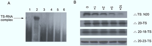
Interaction of TS:N20 with zebrafish TS protein in vitro. A, Gel mobility shift analysis of TS:N20 interaction with TS protein. A 20-nt TS RNA, which included nt 13–32, was synthesized and radio-labeled with 32P. 32P-labeled-TS:N20 RNA was incubated in the absence (lane 1) or presence of 100 ng (lane 2) of pure recombinant zebrafish TS protein. A 32P-labeled 17-nt TS encompassing nt 13–32 with a deletion of the GCU sequence, Δ20-TS RNA, was incubated in the presence of 100 ng of zebrafish TS protein (lane 3). Lanes 4 and 5 represent the other two mutation variants, Δ20–18-TS RNA and Δ20–23-TS RNA, respectively, incubated with 100 ng of pure TS protein. TS:N25 RNA was incubated with 100 ng TS protein in the presence of 100 ng unlabeled full-length TS mRNA (lane 6). Samples were resolved on a non-denaturing 5% acrylamide gel and visualized by autoradiography. The specific RNA–protein complex is indicated (arrow). B, Competition analysis using TS oligomers with TS protein. 32P-labeled full-length TS RNA (105 cpm) was incubated with 200 ng of pure zebrafish recombinant TS protein in the presence of 0–10-fold molar excess competitor, TS:N20 RNA (panel 1), Δ20-TS RNA (panel 2), Δ20–18-TS RNA (panel 3) and Δ20–23-TS RNA (panel 4). The RNA–protein complex was resolved on a non-denaturing 5% acrylamide gel and visualized by autoradiography. |
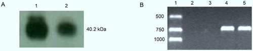
Zebrafish TS protein interacted with its own mRNA in vivo. A, Western blot analysis was performed with purified recombinant zebrafish TS (lane 1) or protein from the immunoprecipitation complex (lane 2) and detected with human TS106 antibody. B, RT-PCR products using the RNA extracted from immunoprecipitation complex as the template. TS mRNA from nt 340–1040 was amplified by RT-PCR using TS-specific primers (lanes 4 and 5). RNA and protein complex was precipitated without TS antibody and RT-PCR was performed (lane 2). Lane 3 indicates the RT-PCR results precipitating the complex with unrelated polyclonal antibody, CHX10-like zebrafish antibody (GenWay Biotech, CA). Lane 1 represents the DNA marker of molecular weight. |
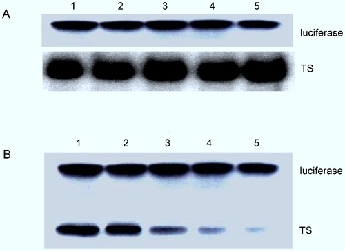
TS protein inhibited in vitro translation of zebrafish TS mRNA. A rabbit reticulocyte lysate in vitro translation system was used to determine the interaction between mRNA and TS protein. Incubation of zebrafish TS mRNA with the reticulocyte lysate yielded a protein product with a molecular mass of approximately 35 kDa. A, Zebrafish TS mRNA was incubated in the rabbit reticulocyte lysate system without (lane 1) or with 100 ng of BSA (lane 2), luciferase (lane 3), human p53 protein (lane 4) or alpha-chymotrypsin (lane 5). B, Excess TS protein was included in the reaction mixture. Zebrafish TS mRNA was incubated in the translation system without (lane 1) or with 1 ng (lane 2), 10 ng (lane 3), 100 ng (lane 4) or 200 ng (lane 5) TS protein. |
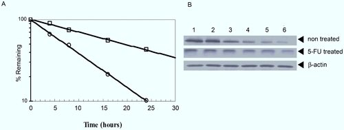
Effect of 5-FU on the stability of TS protein in zebrafish embryos. Healthy embryos at the 1–2 cell stage were treated with 10 μg/mL cycloheximide and 0.5 μg/mL 5-FU and incubated for another 4, 8, 16, 24 and 36 h. Embryos (n = 100) that were untreated or treated with cycloheximide and 5-FU were collected and the total protein was extracted for Western blot analysis as described in the Materials and Methods section. A, TS level in the zebrafish embryo at various time points after 5-FU treatment. TS level in the absence (circles) or presence of 5-FU (box) was analyzed by Western blot and quantified using a Hewlett Packard ScanJet 4P and NIH image 1.51 software. B, Western blot analysis showing the amount of TS at specific times after treatment with cycloheximide. Zebrafish TS embryos were left untreated (panel 1) or treated with 5-FU (panel 2) for 2 h, and 10 μg/mL cycloheximide was added to incubated for another 0 (lane 1), 4 (lane 2), 8 (lane 3), 16 (lane 4), 24 (lane 5) and 36 h (lane 6). Western blots were performed as described in the Materials and Methods section. |