
- Title
-
QDPR homologues in Danio rerio regulate melanin synthesis, early gliogenesis, and glutamine homeostasis
- Authors
- Breuer, M., Guglielmi, L., Zielonka, M., Hemberger, V., K�lker, S., Okun, J.G., Hoffmann, G.F., Carl, M., Sauer, S.W., Opladen, T.
- Source
- Full text @ PLoS One

Characterization of Qdpra. (A) WISH of qdpra at 24 hpf (dorsal view, anterior to the left) shows staining in retinal pigment epithelium (red arrow) and neural crest cells/melanophore precursor (black arrow). At 48 hpf qdpra transcripts are found in the retinal pigment epithelium, choroid fissure (red arrow) and neural crest cells (black arrow). At 72 hpf and more pronounced at 120 hpf (lateral views, anterior to the left), staining is present in the liver (blue arrow). (B) Lateral views with anterior to the left and (C) dorsal views with anterior to the top of embryos at stages indicated. Knockdown of qdpra results in reduced pigments in the eye (asterisk) at 26 hpf (B) and overall diminished pigmentation at 72 hpf (C). (D) At 72 hpf melanin content is reduced by 20% (of wildtype) in Qdpra hypomorphic zebrafish. (E) Amino acid analysis shows hyperphenylalaninemia and normal tyrosine upon qdpraknockdown. PHENOTYPE:
|

Characterization of Qdprb1. (A) WISH of qdprb1 at 24 hpf (dorsal view, anterior to the left) shows staining in eye (red arrow) and mid-hindbrain boundary (black arrow), at 48 hpf (lateral view, anterior to the left) in the optic tectum (blue arrow) and the mid-hindbrain boundary (black arrow) and CMZ (inset; red arrow, dorsal view, anterior to the left), and at 72 hpf (dorsal view, anterior to the left) in proliferative regions of the optic tectum (black arrow, left picture), CMZ (red arrow, right picture, dorsal view of the eye), as well as inner retinal layer (black arrow). (B) Lateral views, anterior to the left. Comparison of wildtype embryos at 18 hpf, 24 hpf and 72 hpf and qdprb1 hypomorphic embryos exhibit abnormal midbrain (red arrow) and anterior hindbrain (blue arrow) morphology and microcephaly. EXPRESSION / LABELING:
PHENOTYPE:
|

ZFIN is incorporating published figure images and captions as part of an ongoing project. Figures from some publications have not yet been curated, or are not available for display because of copyright restrictions. PHENOTYPE:
|
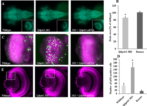
Reduced brain size but increased number of pH3-positive cells upon qdprb1knockdown. (A) Dorsal views, anterior to the left. At 3 dpf tg(HuC/D:GFP) transgenic zebrafish show a decreased size of the optic tectum and eye upon qdprb1 knockdown, which can be rescued by co-injecting qdprb1 mRNA. The insets show dorsal views of the left eye, which is reduced in size but still layered upon qdprb1 suppression. This phenotype is rescued upon co-injection of qdprb1 mRNA. The overall GFP signal remains unchanged. (B) The brain area of Qdprb1 hypormorphic embryos is reduced by about 15% compared to wildtypes and rescued by co-injection of qdprb1 mRNA. (C) Z-stack overlays of DAPI (pink) and pH3 (green) staining of dorsally imaged retinas reveal an increased number of pH3-positive retinal cells in Qdprb1 hypormorphic embryos (3 dpf), which are not restricted to the CMZ like found in wildtype retinas. Number and distribution of pH3-positive cells is restored by co-injection of qdprb1 mRNA. (D) Statistical analysis shows a significant increase in pH3 positive cells upon qdprb1knockdown (n = 6) in comparison to wildtype zebrafish (n = 7), which could be rescued by addition of qdprb1 mRNA (n = 3). (E) Z-confocal image of DAPI staining (pink) of the retina at 3 dpf shows retinal layers although decreased overall size and broadened CMZ in qdprb1 hypomorphic embryos. PHENOTYPE:
|
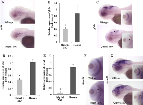
Expression of astroglial markers at 72hpf. (A) Lateral views with anterior to the left. WISH of the BH4 de novo synthesis pathway initiator and dopaminergic neuron marker, gch1, shows unchanged staining upon qdprb1knockdown. (B) RT-qPCR analysis reveals strongly reduced expression of glula in qdprb1 hypomorphic embryos, which is rescued by qdprb1 mRNA co-injection. (C) Lateral views with anterior to the left. This finding is corroborated by WISH experiments highlighting that glula expression is lost in the eye (asterisk in inset) and reduced in the mid/hindbrain (asterisk). (D) Also gfap expression is reduced by qdprb1 knockdown and can be rescued by qdprb1 mRNA co-injection. (E) RT-qPCR analysis of astrocytic glutamate transporters show an almost complete loss of slc1a2a, which is confirmed by WISH (F)–dorsal views focused on the eye. Asterisk indicates the effect on slc1a2aexpression. Slc1a2a expression is normalized in qdprb1 mRNA co-injected embryos (E). (G) Lateral views with anterior to the left. WISH of slc1a2b reveals mildly reduced staining in midbrain and eye (inset), confirming that both SLC1A2 homologues are affected in Qdprb1 hypomorphic embryos. EXPRESSION / LABELING:
PHENOTYPE:
|

Glutamine exposure partially mimics qdprb1 knockdown. (A) Lateral views with anterior to the left. Zebrafish exposed to 20 mM glutamine from sphere stage till 72 hpf display smaller heads and eyes. (B) RT-qPCR analysis zebrafish early exposed to glutamine (20 mM) shows reduced expression of qdprb1, gfap, and slc1a2a. (C) Late exposure to glutamine starting at 48 hpf does not affect the expression of these genes. EXPRESSION / LABELING:
PHENOTYPE:
|
|
ZFIN is incorporating published figure images and captions as part of an ongoing project. Figures from some publications have not yet been curated, or are not available for display because of copyright restrictions. |
|
Characterization of Qdpra.(A) Lateral views, anterior to the left. Expression of Pah at 72 hpf is found in retinal pigment epithelium (red arrow), fin bud (blue arrow) and liver (white arrow). (B) RT-PCR shows loss of exon 3 upon splice blocking MO injection. (C) Lateral views, anterior to the left of 72 hpf embryos. Aberrant pigmentation of Qdpra hypomorphic embryos can be rescued by co-injection of qdpra mRNA. EXPRESSION / LABELING:
|
|
Characterization of Qdprb1.(A) Lateral views, anterior to the left of 72 hpf embryos. p53 knockdown does not rescue the microcephaly phenotype of Qdprb1 hypomorphic embryos. (B) RT-PCR confirms the predicted inclusion of intron 3 upon injection of the splice blocking MO resulting in a strong reduction of correctly spliced mRNA (RT-qPCR, C). (D). Qdprb1 morphant phenotypes using low concentrations of each MO and a combination of both showing a synergy effect between both. (E) Lateral view, 72hpf ATG MO Qdprb1 injected embryos reproduce the phenotype of Splice MO Qdprb1 hypomorphic embryos. (F) Lateral views, anterior to the left. Co-injection of qdprb1mRNA in Qdprb1 hypomorphic embryos rescues brain development. PHENOTYPE:
|
|
ZFIN is incorporating published figure images and captions as part of an ongoing project. Figures from some publications have not yet been curated, or are not available for display because of copyright restrictions. PHENOTYPE:
|
|
Brain development in Qdprb1 hypomorphic embryos.(A) Lateral views, anterior to the left of 72 hpf embryos. Qdprb1 knock down does not affect development of for instance motor neurons (left) and lateral line organ (right) in tg(NBT/lyn:GFP) transgenic zebrafish (red arrows). (B) Lateral views, anterior to the left and (C) dorsal views with anterior to the left at 26 hpf stained for wnt1 (B) and otx2 (C) expression show unchanged expression patterns but reduced size of the positively stained region upon qdprb1knockdown. (D) Z-stacks of DAPI (pink) and pH3 (green) staining of the optic tectum reveals an increase of proliferating cells in 72 hpf Qdprb1 hypomorphic embryos. |
|
WISH and RT-qPCR of glutamate and other solute carrier transporters.RT-qPCR (A) analysis and WISH (B; lateral views, anterior to the left) shows unchanged expression of slc1a3b in Qdprb1 hypomorphic embryos. (C) Further, expression of slc7a5remained unchanged and of slc38a2 was reduced in these zebrafish. EXPRESSION / LABELING:
PHENOTYPE:
|
|
Unillustrated author statements PHENOTYPE:
|