
- Title
-
Capn5 Expression in the Healthy and Regenerating Zebrafish Retina
- Authors
- Coomer, C.E., Morris, A.C.
- Source
- Full text @ Invest. Ophthalmol. Vis. Sci.

capn5a and capn5b are expressed in the developing brain of zebrafish. (A) RT-PCR for capn5a and capn5b expression during development (24, 48, 72, and 120 hpf). atp5h expression is shown as housekeeping gene control. RT, reverse transcriptase; NT, no template. (B) qPCR representation of fold-change in mRNA expression relative to 24 hpf. (C–C″) WISH of capn5a at 18 hpf. Strong capn5a expression was observed in the optic vesicles and brain. WISH with a control sense probe is shown. (C″, D–D″) WISH for capn5a at 24 hpf. Strong expression of capn5a was detected in the brain and hatching gland. Control sense probe is shown. (D″, E–E″) WISH for capn5b at 18 hpf. Expression was observed in parts of the developing brain but not the optic vesicles. Sense probe shown in (E″, F–F″) WISH for capn5b at 24 hpf. Modest expression of capn5b was observed in the brain. c, cerebellum; fp, floor plate; H, hindbrain; HG, hatching gland; OV, optic vesicle; mc, mesencephalon; r, retina; T, telencephalon. Scale bars: 50 μm.
|

Capn5 is expressed in differentiated photoreceptors of the larval zebrafish retina. (A–B′) FISH showing expression of capn5a in the zebrafish retina at 5 dpf. Expression was detected in the ONL (arrows). (B–B′) show an enlarged image of the boxed area in (A–A′). (C–D′) FISH for expression of capn5b in the 5 dpf zebrafish retina. Expression was also detected in the ONL (arrows). (D–D′) show an enlarged image of the boxed area in (C–C′). (E–F′) IHC showing expression of Capn5a/b in the OPL and inner segments of the zebrafish photoreceptors at 5 dpf (arrows). (F–F′) show an enlarged image of the boxed region in (E–E′). GCL, ganglion cell layer; L, lens (the red fluorescence in the lens is nonspecific autofluorescence). Scale bars: 50 μm in (A), (C), and (E) and 100 μm in (B), (D), and (F).
EXPRESSION / LABELING:
|
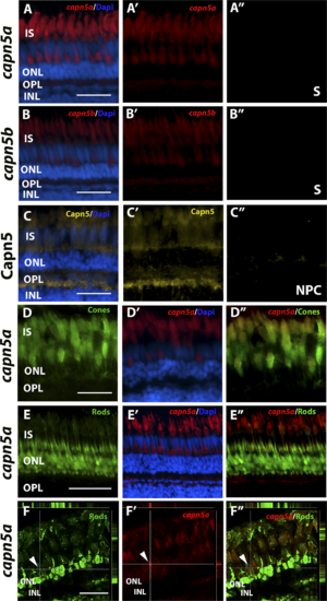
Capn5 is expressed specifically in the cone photoreceptors of the adult zebrafish retina. (A–B″) FISH showing expression of capn5a and capn5b in the adult (WT) retina; expression was seen in the inner segments of the cones. (A″) and (B″) show sense probe controls. (C–C″) IHC for Capn5 (both Capn5a and Capn5b). Expression was observed in the photoreceptor inner segments and in the OPL. (C″) shows control image with no primary antibody. (D–D″) Colocalization of capn5a with a cone-specific marker. Combined FISH for capn5a and IHC for GFP on the TαC:GFP transgenic background demonstrates strong colocalization of capn5a expression with cone-specific GFP expression. (E–E″) Combined FISH for capn5a and IHC for GFP on the XOPS:GFP transgenic background demonstrates a lack of colocalization of capn5a expression with rod-specific GFP expression. (F–F″) Confocal microscopy imaging of combined FISH for capn5a and IHC for GFP on the XOPS:GFP transgenic background; the orthogonal view confirms there is no colocalization of rod-specific GFP and capn5a expression. IS, inner segments; NPC, no primary antibody control; S, sense probe. Scale bars: 50 μm in (E) and 100 μm in (A–D) and (F).
EXPRESSION / LABELING:
|

Expression of Capn5 is elevated in the adult XOPS:mCFP retina. (A) RT-PCR for capn5a and capn5b expression in WT and XOPS:mCFP retinas. Capn5a and capn5b are both expressed in the adult WT retina and capn5a levels are increased in the XOPS:mCFP retina. NT, no template control. (B) qPCR for capn5a and capn5b in WT and XOPS:mCFP adult retina; a 4-fold increase of capn5a expression was observed in the XOPS:mCFP retina compared with WT. (C–C′) FISH showing expression of capn5a in the adult (WT) retina; expression was seen in the IS of the cones. (D–D′) FISH showing expression of capn5a in the XOPS:mCFP retina; expression in the cone IS appears to be more intense than in WT. (E–E′) FISH for capn5b in the adult WT retina; modest expression was detected in the ISs. (F–F′) FISH for capn5b in the XOPS:mCFP retina; expression levels appears similar to WT. (G–G′) IHC for Capn5 (a, b) in the WT adult retina; expression was detected in the OPL and in the cone inner segments (IS). (H–H′) IHC for Capn5 in the XOPS:mCFP retina; increased expression was detected in the OPL and IS. Scale bars: 100 μm. *P < 0.05.
|
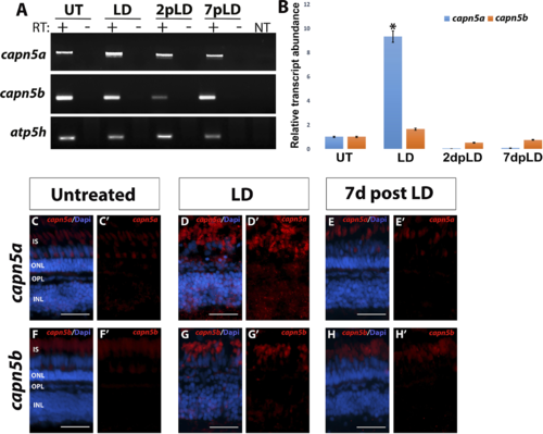
Capn5a expression is induced in response to acute LD. (A) RT-PCR of capn5a and capn5b expression during and following acute LD. A significant increase in expression of capn5a was observed after 3 days of LD, which returned to WT levels by 2 days post LD. Capn5b expression did not change during LD. (B) qPCR for capn5a and capn5b during and following LD. A 12-fold increase in capn5a expression was observed in the LD retina compared with untreated retina, followed by a significant decrease in expression post LD. Capn5b expression did not change during or after LD. (C–E′) FISH for capn5a in untreated retina (UT), during LD, and post LD (7 days post LD). Whereas capn5a expression in the UT retina was confined to the OPL and cone IS, capn5a expression in the LD retina was seen in the INL as well as the OPL and IS. (F–H′) FISH for capn5b in the UT, LD, and 7 days post LD retina. The capn5b expression pattern did not change during or after LD. Scale bars: 50 μm. *P < 0.05.
|
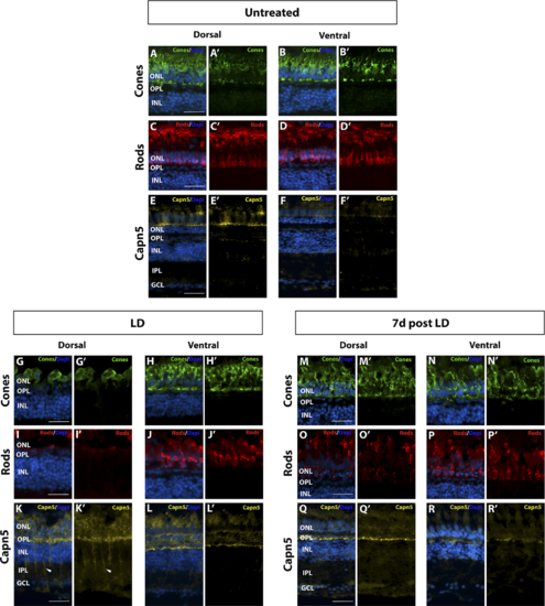
Increase in Capn5 expression correlates with magnitude of retinal damage. (A–B′) Dorsal and ventral expression of cone-specific marker Zpr-1 in undamaged (UT) retina. (C–D′) Dorsal and ventral expression of rod-specific marker 4C12 in the UT retina. (E–F′) IHC for dorsal and ventral Capn5 expression in the UT retina; Capn5 expression is stronger in the dorsal retina (E–E′) compared with the ventral retina (F–F′). (G–H′) Dorsal and ventral cone damage after acute light exposure. There is a significant decrease in the number of cone photoreceptors in the dorsal retina compared with the ventral retina. (I–J′) Dorsal and ventral rod damage after acute light exposure. Rods are almost totally ablated in the dorsal retina, with more moderate damage observed in the ventral retina. (K–L′) IHC for Capn5 expression in the LD retina. Capn5 is strongly upregulated in the INL and surviving cones in the dorsal retina, and more modestly upregulated in the ventral retina. (M–N′) Dorsal and ventral cones have mostly recovered by 7 days post LD. (O–P′) At 7 days post LD, rods are regenerating, with more rods observed in the ventral compared with the dorsal retina. (Q–R′) IHC for Capn5 expression in the 7 days post LD retina. Expression of Capn5 is similar to that of the undamaged retina. Scale bars: 50 μm.
|

capn5a is expressed in the Müller glia in response to acute LD. (A–C′) IHC for Capn5 expression (A) and the Müller glial marker Zrf–1 (B) in the undamaged (UT) adult retina. No colocalization of Capn5 and Müller cells is observed. (A′–C′) show an enlarged image of the boxed regions in (A–C). (D–F) IHC for Capn5 and Müller glia in the LD retina. Capn5 expression is induced in the INL, where it colocalizes with reactive Müller glia (asterisk). (D′–F′) show enlarged images of the boxed regions in (E–F). IPL, inner plexiform layer. Scale bars: 100 μm in (A–F) and 500 μm in (A′–F′).
|

ZFIN is incorporating published figure images and captions as part of an ongoing project. Figures from some publications have not yet been curated, or are not available for display because of copyright restrictions. EXPRESSION / LABELING:
|
|
Unillustrated author statements |