
- Title
-
Arteries define the position of the thyroid gland during its developmental relocalisation
- Authors
- Alt, B., Elsalini, O.A., Schrumpf, P., Haufs, N., Lawson, N.D., Schwabe, G.C., Mundlos, S., Gruters, A., Krude, H., and Rohr, K.B.
- Source
- Full text @ Development
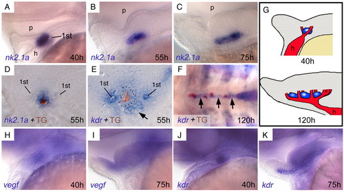
The thyroid develops adjacent to the growing ventral aorta in zebrafish embryos. (A-C) Expression of the thyroid marker nk2.1a (blue) shows primordial expansion along the AP axis around 55 hpf. Lateral views, anterior towards the left. (D) An antibody against thyroglobulin (TG) visualises the lumen of thyroid follicles (brown), surrounded by follicular cells (blue). (E) The endothelial marker kdr is expressed in pharyngeal vessels around the thyroid (lumen brown, broken line around follicle cells), here showing the anterior tip of the ventral aorta on the level of the heart (arrow) and the first pair of branchial arteries (1st). (F) Throughout further development, thyroid tissue (follicles in brown) grows along the ventral aorta (arrows). Ventral view, anterior towards the left. (G) Schematic drawing summarising thyroid and vessel development. (H,I) vegf is diffusely expressed in the ventral pharyngeal area during ventral aorta development. Lateral views, anterior towards the left. (J,K) kdr is diffusely expressed in ventral aorta and surrounding mesenchyme (see also E). Lateral views, anterior towards the left. 1st, first pair of branchial arteries. h, heart; p, pharynx. |
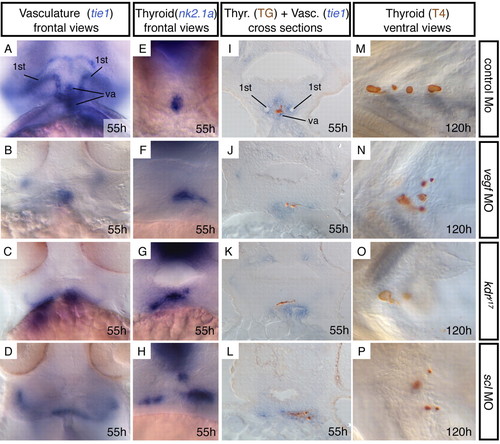
Mutants with defects in ventral aorta development show correlating thyroid abnormalities. (A-H) Frontal views, showing vasculature (tie1 expression) or thyroid (nk2.1a expression). Control morpholino injected embryos are indistinguishable from wild-type embryos or kdry17 siblings. (I-L) Sections showing folliclular lumen (thyroglobulin immunostaining, brown) in relation to endothelial cells (tie1 expression, blue). (M-P) At 120 hpf, thyroid follicles (T4 immunostaining, brown) fail to align along the ventral aorta. In F-H,J-L the thyroid appears larger owing to the lateral expansion. However, in wild-type embryos the thyroid extends along the AP axis (compare with Fig. 1B). Concomitant with lateral expansion, its AP extent appears to be reduced, so that the size remains similar. This is also reflected by normal follicle numbers at 120 hpf (M-P). TG, thyroglobulin; Thyr, thyroid; Vasc, vasculature; va, ventral aorta; 1st, first pair of branchial arteries. EXPRESSION / LABELING:
|
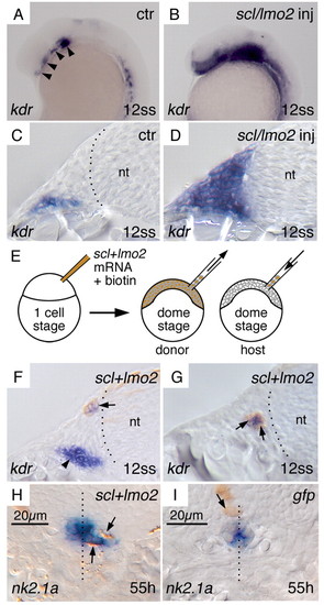
Ectopic endothelial cells can influence thyroid morphology cell non-autonomously. Lateral views (A,B) and cross-sections (C-I) through the head region. (A,C) In wild-type embryos, bilateral groups of kdr domains (arrowheads) mark part of the anterior (head) lateral plate mesoderm that give rise to vascular structures. (B,D) Co-injection of scl and lmo2 mRNA leads to massive upregulation of kdr in the head mesenchyme of zebrafish embryos. (E) Experimental procedure: donor cells from scl and lmo2 co-injected embryos were grafted into wild-type embryos. (F,G) Donor cells have all developed into endothelial cells when found in the head mesenchyme of host embryos. Arrows indicate kdr expression (blue) overlapping with the cell marker biotin (brown) present in grafted cells. Endogenous kdr expression (arrowhead in F) is variably visible on sections, depending on the exact level of sectioning (compare with A). (H) At 55 hpf, grafted scl+lmo2 cells (brown, arrows) have caused the thyroid primordium (blue) to expand laterally. One of the four embryos where grafted cells were detected adjacent to the thyroid primordium. (I) An embryo where gfp injected donor cells are found close to the thyroid, which is not expanded towards the grafted cell. In H and I, care was taken to chose the section through the median level of the thyroid, and diameters were measured on the base of these sections. In I, nuclei in the thyroid appear more prominent than in H, so that the cytoplasmic nk2.1a signal looks slightly weaker on this median section. Broken lines indicate the border of the neural tube (nt) in C,F,G, and the midline in H,I. 12 ss, 12 somite stage. EXPRESSION / LABELING:
|
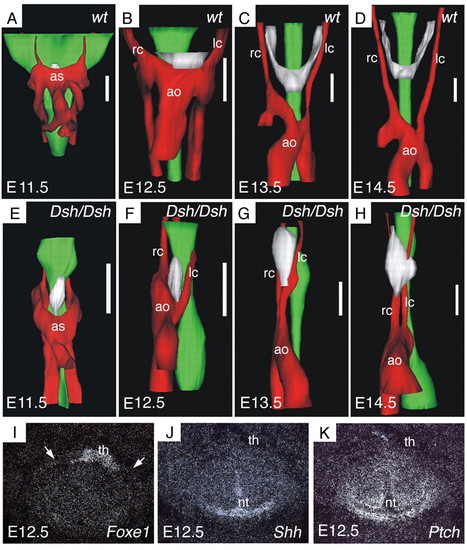
Thyroid morphogenesis is disturbed in short digits (Dsh/Dsh) mutant mice. (A-D) Three-dimensional reconstruction of the cervical region in wild-type mice. (E-H) Same reconstruction of Dsh/Dsh mutant mice. Stages are indicated in the bottom left-hand corner. Red, vascular structures; green, oesophagus; white, thyroid primordium. Scale bar: 140 μm. (I-K) Expression of sonic hedgehog (J) and patched (K) in wild-type E12.5 mouse embryos. Consecutive sections processed for in situ hybridisation, in comparison with the thyroid marker Foxe1 (I). Shh and Ptch are expressed in or around the neural tube, but not in the area of the thyroid. White arrows indicate the position of the carotid arteries (visible in corresponding bright field views). ao, aortic arch; as, aortic sac; lc, left carotid artery; nt, neural tube; rc, right carotid artery; th, thyroid. |
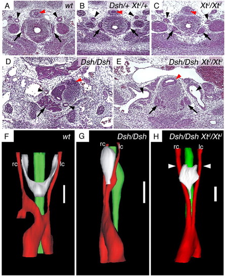
In Dsh/Dsh XtJ/XtJ double mutants, symmetry of carotid arteries is restored and the thyroid primordium regains its midline position. HE staining of representative histological sections (A-E) and three-dimensional reconstruction (F-H) of E13.5 mouse embryos. (A-C) Both carotid arteries (arrowheads) and thyroid lobes (arrows) are normal in double heterozygous or XtJ/XtJ mouse embryos. (D,E) Whereas in Dsh/Dsh mice both carotid arteries and mis-shaped thyroid are positioned unilaterally (D), a bilateral set of carotid arteries (arrowheads) forms in double homozygous Dsh/Dsh XtJ/XtJ mice (E), and the thyroid develops into a bilateral set of lobes at its cranial end (arrows). The carotid arteries in E are larger than in wild type but their identity is unequivocally due to their arterial walls and origin from the aortic arch (as revealed by series of sections, data not shown). Red arrowheads indicate the oesophagus. A-E are at the same magnification; D,E show larger areas. (F-H) Three-dimensional reconstruction visualising how the carotid arteries and the thyroid have regained bilateral positions in Dsh/Dsh XtJ/XtJ mice. White arrowheads indicate the level of the section in E, showing the symmetrical cranial lobes of the thyroid. The thyroid is still abnormally shaped, probably owing to the close distance between the carotid arteries. lc, left carotid artery; rc, right carotid artery. Scale bars: 140 μm. |

Unillustrated author statements EXPRESSION / LABELING:
|