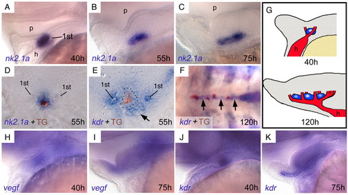
The thyroid develops adjacent to the growing ventral aorta in zebrafish embryos. (A-C) Expression of the thyroid marker nk2.1a (blue) shows primordial expansion along the AP axis around 55 hpf. Lateral views, anterior towards the left. (D) An antibody against thyroglobulin (TG) visualises the lumen of thyroid follicles (brown), surrounded by follicular cells (blue). (E) The endothelial marker kdr is expressed in pharyngeal vessels around the thyroid (lumen brown, broken line around follicle cells), here showing the anterior tip of the ventral aorta on the level of the heart (arrow) and the first pair of branchial arteries (1st). (F) Throughout further development, thyroid tissue (follicles in brown) grows along the ventral aorta (arrows). Ventral view, anterior towards the left. (G) Schematic drawing summarising thyroid and vessel development. (H,I) vegf is diffusely expressed in the ventral pharyngeal area during ventral aorta development. Lateral views, anterior towards the left. (J,K) kdr is diffusely expressed in ventral aorta and surrounding mesenchyme (see also E). Lateral views, anterior towards the left. 1st, first pair of branchial arteries. h, heart; p, pharynx.
|
