IMAGE

Fig 5

ID
ZDB-IMAGE-200612-13
Genes
Source
Figures for Weinschutz Mendes et al., 2020
Image
Figure Caption

Fig 5 Expression of <italic>dlx1a</italic>, <italic>dlx2a</italic>, <italic>dlx5a</italic> and <italic>dlx6a</italic> post-lesion in adult zebrafish.

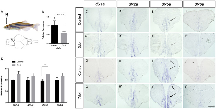
Top left panel (A) shows the location of the mechanical lesion. Expression of the four dlx paralogs in controls (C-F and G-J) and lesioned brains at 3 dpl (C’-F’) and 7 dpl (G’-J’). Down-regulation of dlx5a is suggested at 3dpl (E’ indicated by gray arrow) and confirmed by RT-qPCR (B) (Student’s t-test, n = 5, p = 0.004). A slight up-regulation of dlx2a and dlx5a is seen at 7dpl compared to controls. For each gene, and for each time point, assays were carried out with at least 2 biological replicates in 3 different experimental repetitions (N = 6). RT-qPCR analyses at 7dpl (K) reveals no significant changes in expression levels of dlx1a, dlx2a and dlx6a (N = 7 for each gene each). A significant increase in dlx5a expression was observed at 7dpl (Student’s t-test, n = 7, p = 0.008). Scale bar: 400 μM.

Figure Data
Acknowledgments
This image is the copyrighted work of the attributed author or publisher, and ZFIN has permission only to display this image to its users. Additional permissions should be obtained from the applicable author or publisher of the image. Full text @ PLoS One