
IMAGE
Fig. 4
Image
|
Figure Caption
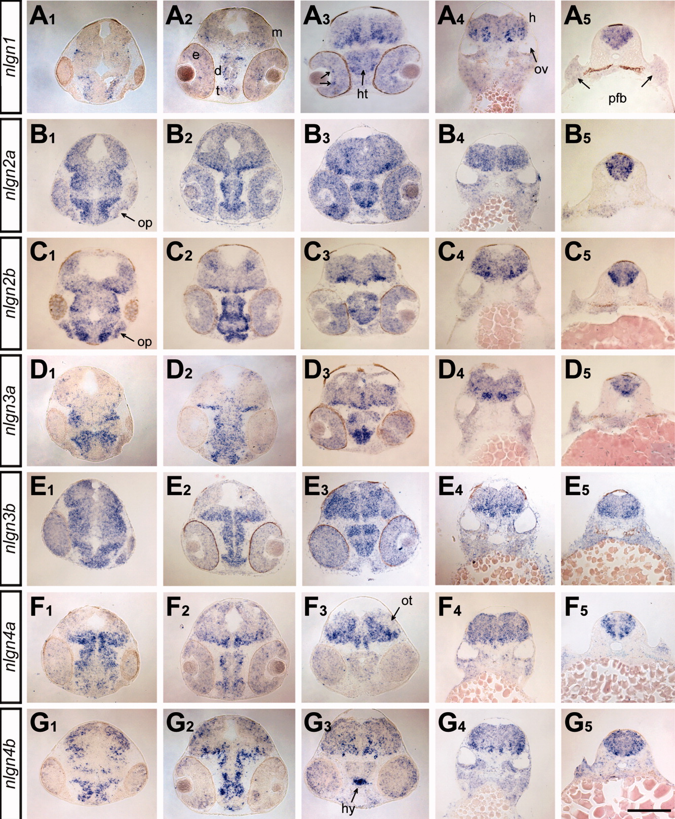
Fig. 4 Expression of nlgns in the developing brain at 48 hpf. The expression patterns for the nlgn genes (A-G) were examined by ISH in cross-sections of embryos at 48 hpf. The levels of the sections (1-5) correspond to sections 2, 3, 5, 8, and 12, respectively, in the ZFIN atlas of zebrafish anatomy (zfin. org/zf_info/anatomy/48hrs/48hrs.html). d, diencephalon; e, eye; m, midbrain; h, hindbrain; ht, hypothalamus; hy, hypophysis; op, olfactory placode; ot, optic tectum; ov, otic vesicle; pfb, pectoral fin bud. Scale bar = 105 μm in A1-G4 and 100 μm in A5-G5.
Figure Data
Acknowledgments
This image is the copyrighted work of the attributed author or publisher, and
ZFIN has permission only to display this image to its users.
Additional permissions should be obtained from the applicable author or publisher of the image.
Full text @ Dev. Dyn.