
Fig. 7
|
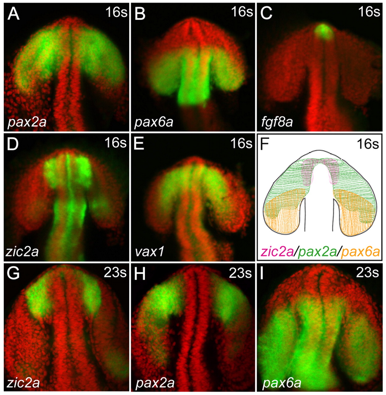
Fig. 7 Patterned gene expression in OS and retinal precursors. (A-F) Wild-type embryos at 16s stained using wholemount in situ hybridization (WISH, green) for expression of pax2a (A), pax6a (B), fgf8a (C), zic2a (D) and vax1 (E). Nuclei were counterstained using DAPI (red). The schematic (F) illustrates the overlap between zic2a and pax2a expression domains in the presumptive OS, and the overlap between pax2a and pax6a domains in the presumptive retina. (G-I) Expression of zic2a (G), pax2a (H) and pax6a (I) detected at 23s. Expression patterns, imaged using confocal microscopy, are shown as z-stacks in ventral view, anterior up.